Canine Anal / Perianal
Sac Tumor
& Breeds at Risk
Research, Resources & Education
This website is based on research and is NOT created to diagnose your pet.
Each animal is an individual and may exhibit symptoms in a different way.
It is advised that you ALWAYS CHECK WITH YOUR VETERINARIAN for a proper diagnosis and treatment plan.
Please visit Lost Temple Fitness & Cancer for more information of cancer in humans including
What is Cancer and Treatments.
Table of Contents
Canine Anal / Perianal Sac Tumor
An anal sac tumor is a tumor made up of cells originating from the glands of the anal sac.
Symptoms may include an external swelling in the perianal region, a mass may be felt during a rectal exam, constipation, pain or straining to defecate, blood in the stool and/or excessive thirst and urination.
Treatments may include chemotherapy and/or radiation treatment, possible removal of lymph nodes if needed and/or palliative care.
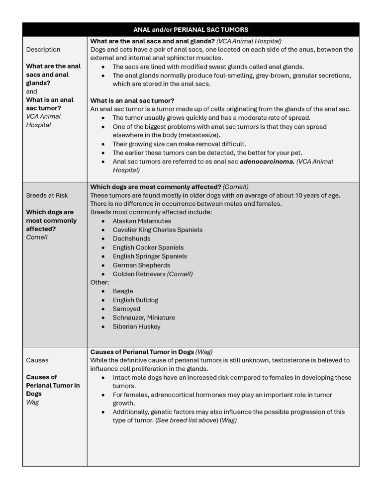
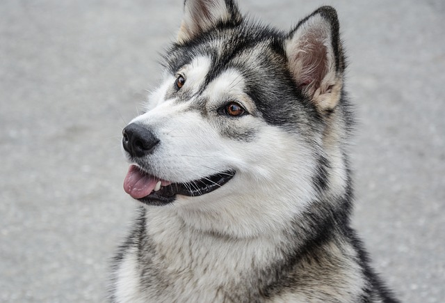
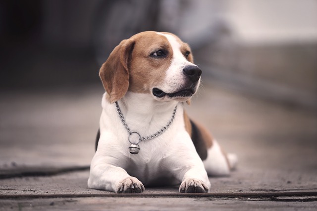
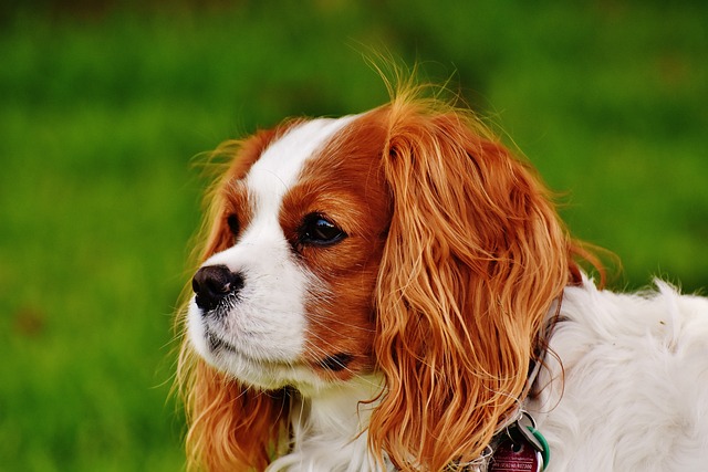
What Dogs are at Risk?
Some Dogs that are at Risk due to Genetic Predisposition

What are Anal or Perianal Sac Tumors
What are the Anal Sacs, Anal Glands and Tumors? (VCA Animal Hospital)
What are the anal sacs and anal glands? (VCA Animal Hospital)
Dogs and cats have a pair of anal sacs, one located on each side of the anus, between the external and internal anal sphincter muscles.
- The sacs are lined with modified sweat glands called anal glands.
- The anal glands normally produce foul-smelling, grey-brown, granular secretions, which are stored in the anal sacs.
What is an anal sac tumor?
An anal sac tumor is a tumor made up of cells originating from the glands of the anal sac.
- The tumor usually grows quickly and has a moderate rate of spread.
- One of the biggest problems with anal sac tumors is that they can spread elsewhere in the body (metastasize).
- Their growing size can make removal difficult.
- The earlier these tumors can be detected, the better for your pet.
- Anal sac tumors are referred to as anal sac (VCA Animal Hospital)
Perianal / Anal Tumor Videos
YouTube Videos that help explain Anal and/or Perianal Gland Tumors in Dogs
Disclaimer:
This is for research only and Lost Temple Pets does not endorse any video presented on this website.
It is advised that you ALWAYS CHECK WITH YOUR VETERINARIAN for a proper diagnosis and treatment plan.
Playlist
Causes & Breeds Commonly Affected
Causes of Perianal Tumor in Dogs (Wag)
Causes of Perianal Tumor in Dogs (Wag)
While the definitive cause of perianal tumors is still unknown, testosterone is believed to influence cell proliferation in the glands.
- Intact male dogs have an increased risk compared to females in developing these tumors.
- For females, adrenocortical hormones may play an important role in tumor growth.
- Additionally, genetic factors may also influence the possible progression of this type of tumor. (See breed list above) (Wag)
Which Dogs are Most Commonly Affected? (Cornell)
Which Dogs are Most Commonly Affected? (Cornell)
These tumors are found mostly in older dogs with an average of about 10 years of age.
Symptoms
Signs & Symptoms (ACVS)
Signs & Symptoms (ACVS)
The signs of anal sac tumors can be variable:
- An external swelling in the perianal region
- A mass may be felt during a routine rectal examination
- Constipation
- Pain or straining to defecate
- Blood in the stool
- Excessive thirst and urination may occur if kidney failure is present from elevated calcium levels
- Without enlarged lymph nodes (in the abdomen), even a large tumor often produces limited symptoms associated with defecation
Animal owners may observe symptoms of kidney failure from the elevated calcium (referred to as hypercalcemia of malignancy).
These symptoms generally include:
- Increased thirst
- increase in urination
- Vomiting
- Loss of appetite
- Lethargy (weakness/tiredness) (ACVS)
Testing / Diagnostics
Diagnostics (ACVS)
Diagnostics (ACVS)
In general, the following tests are recommended to diagnose the tumor, provide a clear clinical picture of overall health and evaluate for metastasis:
- Aspiration: A small needle is inserted into the tumor to obtain a few cells that can differentiate cancer from infection or inflammation.
- Blood tests: Assess overall health. Evaluating for hypercalcemia and kidney failure
- Chest x-rays: Evaluate for metastatic nodules and other heart and lung problems
- Abdominal ultrasound: Examination to evaluate for enlarged lymph nodes or tumor spread into other organs such as liver, kidneys, etc. These enlarged lymph nodes are often what produce symptoms associated with defecation. (ACVS)
Diagnosis of Anal Sac Disease in Dogs and Cats (Merck Manual)
Diagnosis of Anal Sac Disease in Dogs and Cats (Merck Manual)
Physical examination, although microscopy, ultrasonography, or biopsy may be required
- Diagnosis of impaction, infection, or abscessation is confirmed by digital rectal examination, at which time the sacs can be expressed.
- Microscopic examination of the contents from infected sacs reveals large numbers of polymorphonuclear leukocytes and bacteria.
- A tumor should be suspected (anal sac apocrine adenocarcinoma) in anal sacs that are firm, enlarged, and non-expressible even with irrigation.
- Ultrasonography may be useful to determine whether a firm, non-expressible anal sac is due to infection/abscessation or neoplasia.
- In the case of a suspected tumor, the diagnosis should be confirmed by biopsy.
- Regional and systemic metastasis should be evaluated, and serum calcium should be measured. (Merck Manual)
Treatment
Treatment (ACVS)
Treatment (ACVS)
Consultation with your primary care veterinarian may result in a referral to a veterinary surgeon to fully explore your options.
- Surgery is the mainstay of treatment. It is the only proven method to influence survival of dogs with these tumors.
- The tumor is removed through an incision adjacent to the anal opening directly over the tumor.
- Wide and aggressive removal is not feasible due to the adjacent rectum and anus.
- With large tumors, additional tissue attached to the tumor may need to be removed. This may result in some of the complications.
- If there are enlarged lymph nodes in the abdomen, they are removed through an abdominal surgical approach on the underside of the dog.
- These nodes are enlarged in about 50% of cases.
- This can be done at the time of the primary tumor removal, shortly thereafter, or later if these nodes enlarge.
- This procedure is done to alleviate the constipation and difficulty defecating.
- If kidney failure or hypercalcemia is present, therapy with intravenous fluids and medications may be needed prior to surgery to make your dog a more suitable candidate for anesthesia. In some cases, kidney failure can be permanent.
- After surgery, chemotherapy and/or radiation treatment may improve the life expectancy of your pet. (ACVS)
How is it Treated? (Cornell)
How is it Treated? (Cornell)
Dogs with hypercalcemia may need to be stabilized with intravenous fluids and medications, such as corticosteroids or diuretics.
- Surgery to remove the mass and the entire anal sac (anal sacculectomy) is the treatment of choice. Complete removal can be challenging for larger masses due to their proximity to the rectum. One of the major complications of surgery is fecal incontinence, which may be temporary or permanent. Other complications include infection, poor wound healing and anal stricture.
- If enlarged lymph nodes are present, they can be removed via a separate approach through the abdominal wall. This can be a complicated procedure, and it carries risks of severe hemorrhage and nerve damage that can result in temporary or permanent urinary incontinence. The overall complication rate is approximately 10%.
- Radiation therapy may be used preoperatively, intraoperatively or postoperatively on the anal sac mass and the lymph nodes — reducing the risk of recurrence in cases where complete removal is not possible. It can also be used palliatively in cases where surgery is not an option. Complications, including life-threatening ones, can occur.
- Chemotherapy has not been proven to improve survival, but may provide some benefits, especially when combined with radiation therapy.
- Palliative care will include pain medications, such as nonsteroidal anti-inflammatories (NSAIDS), corticosteroids (not if NSAIDS are being used), fluid therapy and diuretics. (Cornell)
Chemotherapy (Improve Vet Practice)
Chemotherapy (Improve Vet Practice)
Chemotherapy, targeted therapy, RT (radiation therapy) or combinations of these treatment modalities can be considered in cases where surgical management is not possible or not elected by the owner, or in cases of stage IV disease.
- The two main chemotherapy drugs with demonstrated efficacy in the gross disease setting include carboplatin (33 percent partial response rate) and actinomycin-D (50 percent partial response rate), although it should be noted that these figures come from very small study populations (Bennett et al., 2002; Hammer et al., 1994).
- Targeted therapy with toceranib phosphate (Palladia) is perhaps a more promising medical option for the management of gross disease, with clinical benefit seen in 69 to 88 percent of cases (Heaton et al., 2020; London et al., 2012); around 20 to 25 percent of dogs will demonstrate a partial response with 60 to 70 percent achieving stable disease.
- A clinical benefit is also observed in the majority of stage IV patients, despite no partial or complete responses being observed in one study (Elliott, 2019).
- Toceranib inhibits a number of targets including VEGFR-2, PDGFR-ß, KIT, FLT3 and CSF1R, a number of which are known to be expressed in canine AGASAC as discussed earlier (Urie et al., 2012).
- Regarding RT, in the gross disease setting response rates of up to 75 percent have been reported, and resolution of hypercalcaemia has been reported in 31 percent of cases (McDonald et al., 2012).
- A variety of protocols, both definitive-intent and palliative-intent, have been described and can be tailored to patient and client needs. In some specific situations, such as stage IIIb disease, RT may be superior to surgery (Meier et al., 2017). (Improve Vet Practice)
Follow up & Prognosis
Aftercare and Outcome (ACVS)
Aftercare and Outcome (ACVS)
Most animals are discharged 1-2 days after surgery.
- There is usually a follow-up appointment to see how your dog is doing and to remove skin sutures or staples (if present).
- Pain can be well-controlled with owner-administered medications.
Restrictions following surgery usually are:
- Use of a restrictive collar for 10-14 days after surgery to prevent the natural tendency of dogs to lick and chew at a wound. This can cause breakdown of the wound and infection.
- Stool softening medications may be needed until swelling resolves
- Limited and restricted activity is indicated for about 2 weeks to allow recovery and incision healing
Postoperative complications can include:
- Incision infection
- Wound breakdown (dehiscence)
- Fecal incontinence can occur in up to 33% of dogs especially with removal of larger masses. This is usually temporary, but owners need to be aware of this problem.
- If the tumor is only on one side, the incontinence is typically partial in that the dog has difficulty controlling bowel movements but not continuous dropping of stool
- Continued kidney problems
The prognosis with apocrine gland adenocarcinoma depends on type of treatment, size of mass, presence of hypercalcemia and presence of lymph node involvement.
- Surgical removal of these nodes can produce long-term relief of constipation.
- Some animals have had multiple surgeries to remove recurrent lymph nodes to alleviate obstructions successfully.
It is important that your veterinarian examines the anal sacs as part of your dog’s routine examination. Early detection can greatly improve survival. (ACVS)
References
References
ACVS – College of Vet surgeons – Anal Sac Tumors in Dogs
https://www.acvs.org/small-animal/anal-sac-tumors-in-dogs/
Cornell Richard P. Riney Canine Health Center – Anal sac adenocarcinoma
Improve Vet Practice – Diagnosis and management of canine anal sac adenocarcinomas
by Andy Yale
02 September 2022
PUBLISHED IN: DOGONCOLOGYSMALL ANIMALVET
Merck Manual – Anal Sac Disease in Dogs and Cats
By Alex Gallagher, DVM, MS, DACVIM-SAIM
Reviewed/Revised Oct 2020
VCA Animal Hospital
Anal Sac Tumors
By Malcolm Weir, DVM, MSc, MPH; Christopher Pinard, DVM, DVSc, DACVIM (Oncology)
https://vcahospitals.com/know-your-pet/anal-gland-tumors
Wag – Perianal Tumor in Dogs
Written By Grace Park, Published: 04/06/2017Updated: 04/05/2021
Veterinary reviewed by Michele K.
https://wagwalking.com/condition/perianal-gland-tumor
Cancer/Breed Chart
| BREED | BRAIN | HEMANGIO- SARCOMA | LYMPHOMA | MAMMARY TUMORS | MAST CELL TUMOR | MELANOMA | NASAL TUMOR | ORAL | OSTEOSARCOMA | PERIANAL/ ANAL SAC | SOFT TISSUE SARCOMA | TRANSITIONAL (TCC) / UROTHELIAL (UC) |
|---|---|---|---|---|---|---|---|---|---|---|---|---|
| Airedale Terrier | Lymphoma | Melanoma | Nasal Tumor | Soft Tissue Sarcoma | TCC or UC | |||||||
| Basset Hound | Hemangiosarcoma | Lymphoma | Nasal Tumor | Soft Tissue Sarcoma | ||||||||
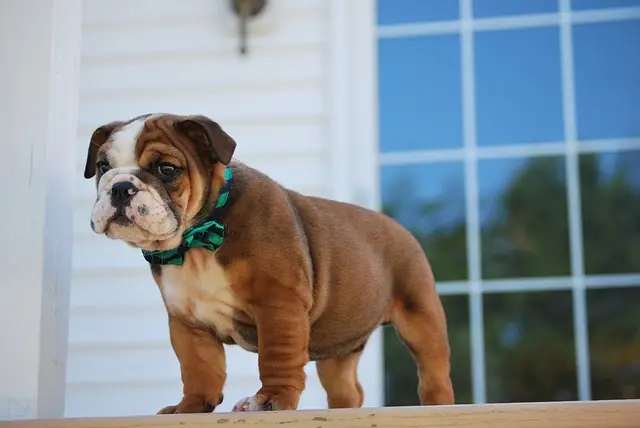
| Bulldog, English | Brain | Lymphoma | Mast Cell Tumor | Perianal/Anal Sac | Soft Tissue Sarcoma | |||||||
| Bullmastiff | Lymphoma | Mast Cell Tumor | Soft Tissue Sarcoma | |||||||||
| St. Bernard | Lymphoma | Osteosarcoma | Soft Tissue Sarcoma | |||||||||
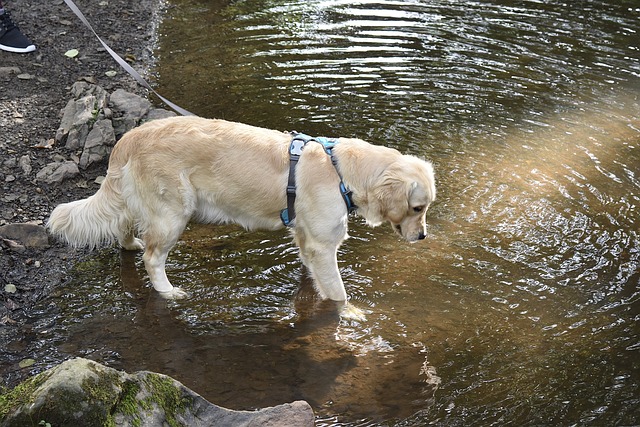
| Golden Retriever | Brain | Hemangiosarcoma | Lymphoma | Mast Cell Tumor | Melanoma | Oral | Osteosarcoma | Perianal/Anal Sac | Soft Tissue Sarcoma | |||
| Labrador Retriever | Hemangiosarcoma | Lymphoma | Mast Cell Tumor | Melanoma | Nasal Tumor | Oral | ||||||
| Scottish Terrier | Brain | Lymphoma | Melanoma | Nasal Tumor | Oral | Soft Tissue Sarcoma | TCC or UC | |||||
| Boxer | Brain (Glioma) | Hemangiosarcoma | Lymphoma | Mammary Tumor | Mast Cell Tumor | Oral | Osteosarcoma | Soft Tissue Sarcoma | ||||
| Beagle | Hemangiosarcoma | Lymphoma | Mast Cell Tumor | Perianal/Anal Sac | TCC or UC | |||||||
| West Highland White Terrier | Lymphoma | |||||||||||
| Chow Chow | Lymphoma | Melanoma | Oral | |||||||||
| Poodle, Standard | Lymphoma | Melanoma | Nasal Tumor | Oral | ||||||||
| Rottweiler | Lymphoma | Oral | Osteosarcoma | |||||||||
| Poodle, Toy | Lymphoma | Mammary Tumor | Melanoma | Nasal Tumor | ||||||||
| Yorkshire Terrier | Lymphoma | Mammary Tumor | ||||||||||
| German Shepherd | Hemangiosarcoma | Lymphoma | Mammary Tumor | Melanoma | Nasal Tumor | Oral | Osteosarcoma | Perianal/Anal Sac | ||||
| Poodle, Miniature | Lymphoma | Mammary Tumor | Melanoma | Nasal Tumor | Oral | |||||||
| Affenpinscher | ||||||||||||
| Afghan Hound | ||||||||||||
| Alaskan Malamute | Perianal/Anal Sac | |||||||||||
| American Eskimo, Toy and Standard | ||||||||||||
| American Foxhound | ||||||||||||
| American Pitt Bull Terrier | Hemangiosarcoma | |||||||||||
| American Staffordshire Terrier | ||||||||||||
| American Water Spaniel | ||||||||||||
| Anatolian Shepherd Dog | ||||||||||||
| Australian Cattle Dog | TCC or UC | |||||||||||
| Australian Shepherd | TCC or UC | |||||||||||
| Australian Terrier | ||||||||||||
| Basenji | ||||||||||||
| Bearded Collie | ||||||||||||
| Beauceron | ||||||||||||
| Bedlington Terrier | ||||||||||||
| Belgian Groenendael | ||||||||||||
| Belgian Malinois | ||||||||||||
| Belgian Tervuren | ||||||||||||
| Bernese Mountain Dog | Hemangiosarcoma | Melanoma | Soft Tissue Sarcoma | |||||||||
| Bichon Frise’ | TCC or UC | |||||||||||
| Black and Tan Coonhound | ||||||||||||
| Black Russian Terrier | ||||||||||||
| Bloodhound | Soft Tissue Sarcoma | |||||||||||
| Boerboel | ||||||||||||
| Border Collie | Brain | TCC or UC | ||||||||||
| Border Terrier | ||||||||||||
| Borzoi | Osteosarcoma | |||||||||||
| Boston Terrier | Brain | Mast Cell Tumor | Melanoma | Soft Tissue Sarcoma | ||||||||
| Bouvier des Flandres | Soft Tissue Sarcoma | |||||||||||
| Briard | ||||||||||||
| Brussels Griffon | ||||||||||||
| Bull Terrier | Mast Cell Tumor | Melanoma | ||||||||||
| Bull Terrier, Miniature | Mast Cell Tumor | Melanoma | ||||||||||
| Cairn Terrier | ||||||||||||
| Canaan Dog | ||||||||||||
| Cane Corso (Italian Mastiff) | ||||||||||||
| Caucasian Shepherd | ||||||||||||
| Cavalier King Charles Spaniel | Perianal/Anal Sac | |||||||||||
| Chesapeake Bay Retriever | Melanoma | |||||||||||
| Chinese Crested | ||||||||||||
| Chinese Shar-Pei | Mast Cell Tumor | Soft Tissue Sarcoma | TCC or UC | |||||||||
| Clumber Spaniel | ||||||||||||
| Curly Coated Retriever | ||||||||||||
| Dalmation | Hemangiosarcoma | |||||||||||
| Dandie Dinmont Terrier | ||||||||||||
| Dogo Argentino | ||||||||||||
| Dogue de Bordeaux | ||||||||||||
| English Foxhound | ||||||||||||
| English Toy Spaniel AKA King Charles Spaniel | ||||||||||||
| Field Spaniel | ||||||||||||
| Finnish Spitz | ||||||||||||
| Flat-Coated Retriever | Hemangiosarcoma | |||||||||||
| Fox Terrier, Smooth | Mast Cell Tumor | |||||||||||
| Fox Terrier, Toy | Mast Cell Tumor | |||||||||||
| Fox Terrier, Wire | TCC or UC | |||||||||||
| French Bulldog | ||||||||||||
| German Pinscher | ||||||||||||
| German Wirehaired Pointer | ||||||||||||
| Glen of Imaal Terrier | ||||||||||||
| Great Dane | Brain | Osteosarcoma | Soft Tissue Sarcoma | |||||||||
| Great Pyrenees | ||||||||||||
| Greater Swiss Mountain Dog | ||||||||||||
| Greyhound | Brain (Meningioma) | Hemangiosarcoma | Osteosarcoma | Soft Tissue Sarcoma | ||||||||
| Harrier | ||||||||||||
| Havanese | ||||||||||||
| Ibizan Hound | ||||||||||||
| Irish Terrier | ||||||||||||
| Irish Water Spaniel | ||||||||||||
| Irish Wolfhound | Osteosarcoma | |||||||||||
| Italian Greyhound | Brain | Hemangiosarcoma | ||||||||||
| Japanese Chin | ||||||||||||
| Keeshond | Nasal Tumor | |||||||||||
| Kerry Blue Terrier | ||||||||||||
| Komondor | ||||||||||||
| Kuvasz | ||||||||||||
| Lakeland Terrier | ||||||||||||
| Leonberger | Osteosarcoma | |||||||||||
| Lhasa Apso | TCC or UC | |||||||||||
| Lowchen | ||||||||||||
| Manchester Terrier Toy | ||||||||||||
| Manchester Terrier, Standard | ||||||||||||
| Mastiff | Brain | |||||||||||
| Miniature Pincher | ||||||||||||
| Neapolitan Mastiff | ||||||||||||
| Newfoundland | ||||||||||||
| Norfolk Terrier | ||||||||||||
| Norwegian Buhund | ||||||||||||
| Norwegian Elkhound | Brain | Nasal Tumor | ||||||||||
| Norwich Terrier | ||||||||||||
| Nova Scotia Duck Tolling Retriever | ||||||||||||
| Old English Sheepdog | Brain | |||||||||||
| Otterhound | ||||||||||||
| Papillon | ||||||||||||
| Parsons Russell Terrier | TCC or UC | |||||||||||
| Pekingese | Brain | |||||||||||
| Petit Basset Griffon Vendeen (PBGV) | ||||||||||||
| Pharaoh Hound | ||||||||||||
| Plott Hound | ||||||||||||
| Polish Lowland Sheepdog | ||||||||||||
| Pomeranian | ||||||||||||
| Portuguese Water Dog | Brain | Hemangiosarcoma | ||||||||||
| Presa Canario | ||||||||||||
| Pug | Brain | Mast Cell Tumor | Soft Tissue Sarcoma | |||||||||
| Puli | ||||||||||||
| Pyrenean Shepherd | ||||||||||||
| Rhodesian Ridgeback | Mast Cell Tumor | Soft Tissue Sarcoma | ||||||||||
| Saluki | ||||||||||||
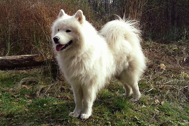
| Samoyed | Perianal/Anal Sac | |||||||||||
| Schipperke | ||||||||||||
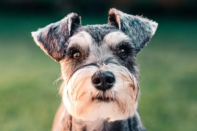
| Schnauzer, Miniature | Mast Cell Tumor | Melanoma | Perianal/Anal Sac | Soft Tissue Sarcoma | ||||||||
| Schnauzer, Standard | Mast Cell Tumor | Melanoma | Soft Tissue Sarcoma | |||||||||
| Sealyham Terrier | ||||||||||||
| Shiba Inu | ||||||||||||
| Shih Tzu | Brain | |||||||||||
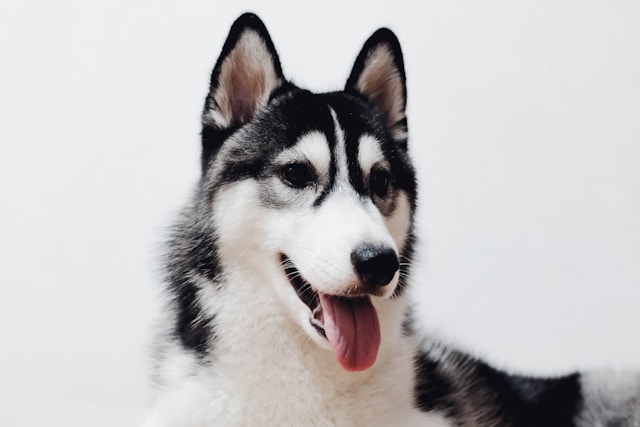
| Siberian Husky | Perianal/Anal Sac | |||||||||||
| Silky Terrier | ||||||||||||
| Skye Terrier | Hemangiosarcoma | |||||||||||
| Soft-Coated Wheaten Terrier | ||||||||||||
| Spinone Italiano | ||||||||||||
| Staffordshire Bull Terrier | Mast Cell Tumor | |||||||||||
| Sussex Spaniel | ||||||||||||
| Swedish Vallhund | ||||||||||||
| Tibetan Mastiff | ||||||||||||
| Tibetan Spaniel | ||||||||||||
| Tibetan Terrier | ||||||||||||
| Tosa | ||||||||||||
| Vizsla | Melanoma | |||||||||||
| Weimaraner | Mast Cell Tumor | |||||||||||
| Welsh Corgi, Cardigan | ||||||||||||
| Welsh Corgi, Pembroke | ||||||||||||
| Welsh Springer Spaniel | ||||||||||||
| Welsh Terrier | ||||||||||||
| Whippet | Hemangiosarcoma | TCC or UC | ||||||||||
| Wirehaired Pointing Griffon | ||||||||||||
| Akita (American) | Oral | |||||||||||
| Collie, Rough / Smooth Coat | Brain (Meningioma) | Nasal Tumor | Oral | TCC or UC | ||||||||
| Gordon Setter | Melanoma | Oral | ||||||||||
| Irish Setter | Melanoma | Oral | Osteosarcoma | Soft Tissue Sarcoma | ||||||||
| Schnauzer, Giant | Mast Cell Tumor | Melanoma | Oral | |||||||||
| Scottish Deerhound | Brain | Melanoma | Oral | Osteosarcoma | TCC or UC | |||||||
| Shetland Sheepdog | Nasal Tumor | Oral | TCC or UC | |||||||||
| Brittany | Mammary Tumor | |||||||||||
| Chihuahua | Mammary Tumor | Melanoma | ||||||||||
| English Cocker Spaniel | Mammary Tumor | |||||||||||
| English Setter | Mammary Tumor | |||||||||||
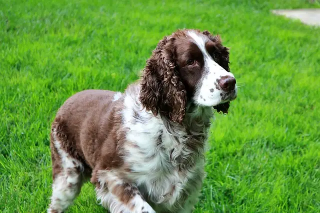
| English Springer Spaniel | Mammary Tumor | Melanoma | Perianal/Anal Sac | |||||||||
| Maltese | Mammary Tumor | |||||||||||
| Pointer | Hemangiosarcoma | Mammary Tumor | ||||||||||
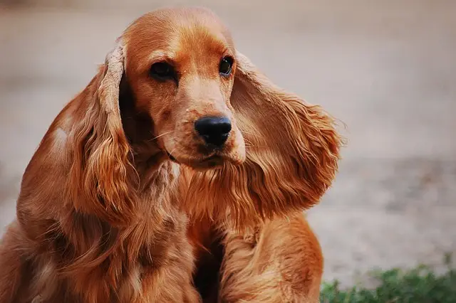
| Cocker Spaniel (American) | Mammary Tumor | Mast Cell Tumor | Melanoma | Oral | Perianal/Anal Sac | |||||||
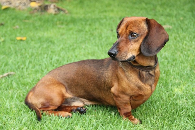
| Dachshund | Brain | Mammary Tumor | Mast Cell Tumor | Oral | Perianal/Anal Sac | |||||||
| Doberman Pinscher | Brain | Mammary Tumor | Melanoma | Oral | Osteosarcoma | |||||||
| German Shorthaired Pointer | Mammary Tumor | Nasal Tumor | Oral |