Canine
Mast Cell Tumors
& Breeds at Risk
Research, Resources & Education
This website is based on research and is NOT created to diagnose your pet.
Each animal is an individual and may exhibit symptoms in a different way.
It is advised that you ALWAYS CHECK WITH YOUR VETERINARIAN for a proper diagnosis and treatment plan.
Please visit Lost Temple Fitness & Cancer for more information of cancer in humans including
What is Cancer and Treatments.
Table of Contents
Canine Mast Cell Tumors
Mast cells are a type of white blood cell that plays a role in the allergic response, which can become cancerous masses in dogs. Tumors are commonly found on the skin or layer under the skin.
Symptoms include enlarged lymph nodes, GI upset, decreased appetite, low energy and/or increased respiratory rate.
Treatments can include radiation, chemotherapy, palliative therapy or an oral medication such as Palladia.
What Dogs are at Risk?
Some Dogs that are at Risk due to Genetic Predisposition


What are Mast Cell Tumors?
What is a Mast Cell and What are Mast Cell Tumors? (VCA Animal Hospitals)
What is a Mast Cell? (VCA Animal Hospitals)
A mast cell is a type of white blood cell that is found in many tissues of the body.
- Mast cells are allergy cells and play a role in the allergic response. When exposed to allergens, mast cells release chemicals and compounds (called degranulation).
- One of these compounds is histamine. Histamine is commonly known for causing itchiness, sneezing, and runny eyes and nose – the common symptoms of allergies.
- When histamine and the other compounds are released in excessive amounts (with mass degranulation), it can cause full-body effects, including anaphylaxis, a serious, life-threatening, allergic reaction.
- Other complications include delayed wound healing, bleeding disorders, and gastrointestinal (GI) ulceration.
What is a mast cell tumor?
A mast cell tumor (MCT) is a type of malignant (cancerous) tumor consisting of mast cells.
- Mast cell tumors typically form nodules or masses in the skin, but they can also affect other areas of the body, including the spleen, liver, intestine, and bone marrow.
- MCTs are the most common skin tumor in dogs (7%–21%).
- Most dogs with MCT (approximately 85%) only develop one tumor. (VCA Animal Hospitals)
Mast Cell Tumors in Dogs (NC State Veterinary Hospital)
Mast Cell Tumors in Dogs (NC State Veterinary Hospital)
Mast cell tumors vary in appearance. Some may look like raised bumps within, or just below the surface of, the skin. Others appear as red, ulcerated, bleeding, bruised, and/or swollen growths.
- Some tumors appear and remain the same size for months or years, while others show a rapid growth pattern over days or weeks.
- They can also increase and decrease in size over time.
Tumors can be irritating, and dogs will scratch, lick, or bite the mass and surrounding skin.
- This trauma causes the tumor cells to release the chemicals in their granules leading to a localized reaction.
- In more serious cases, the chemicals can affect the entire body, causing severe gastrointestinal bleeding and even an anaphylactic reaction, which can be fatal if untreated. (NC State Veterinary Hospital)
Common Locations in the Dog (ACVS)
Common locations in the dog (ACVS) include:
- Trunk 42–65%
- Limbs 22–43%
- Head and neck 10–14%
Visceral (intra-abdominal organs) mast cell disease is a recognized form of the disease and is more aggressive than the aforementioned locations.
- This is often preceded by skin or subcutaneous tumors. (ACVS)
Mast Cell Tumor Videos
YouTube Videos that help explain Mast Cell Tumors in Dogs
Disclaimer:
This is for research only and Lost Temple Pets does not endorse any video presented on this website.
It is advised that you ALWAYS CHECK WITH YOUR VETERINARIAN for a proper diagnosis and treatment plan.
Playlist
Breeds at Risk, Causes and Genetics
Breeds at Risk - Overview (ACVS)
Overview (ACVS)
Mast cell tumors are the most common type of skin tumor found in dogs and the second most common skin tumor in cats. These represent 14–21% of all skin tumors diagnosed in dogs. They are usually noticed in middle aged patients but can occur in patients of any age. Boxers and Boston terriers make up ~ 50% of all cases.
Most tumors are solitary although boxers and pugs have an increased predilection for multiple skin tumors. These tumors most often are noticed in the skin and subcutaneous tissue. (ACVS)
Breeds Prone to Mast Cell Tumors (Better Pet)
Breeds prone to mast cell tumors (Better Pet)
While any breed of dog can get mast cell tumors, certain breeds, especially brachycephalic breeds, are more susceptible. Mast cell tumors are particularly common in:
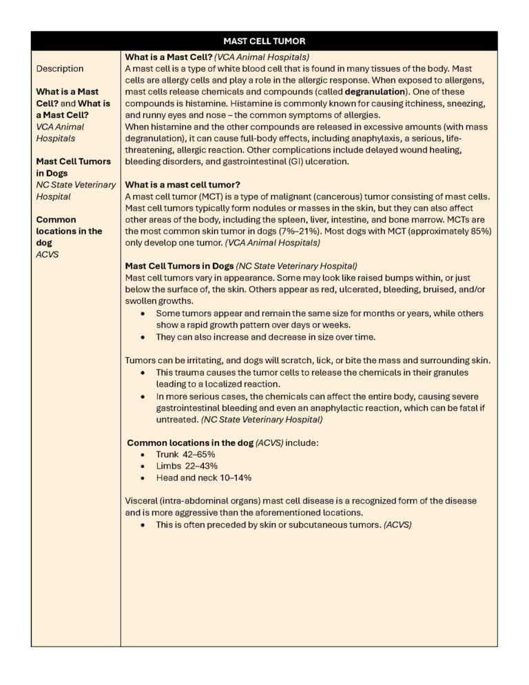
- Boxers. No one knows exactly why boxers are more prone to mast cell tumors, but they do tend to get them more frequently than other breeds. The good news is that their tumors are often (but not always) less severe.
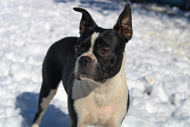
- Bull terriers. The bull terrier is another breed of dog that is predisposed to mast cell tumors. If you have a bull terrier, make sure they are screened for mast cell tumors regularly and that you know the signs of complications from mast cell tumors.
- Boston terriers. Similar to boxers, this is a breed that is prone to getting mast cell tumors. The reason for this is not known.
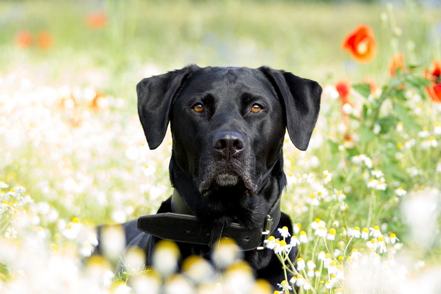
- Labrador retrievers. Labrador retrievers are another breed that is prone to mast cell tumors and particularly aggressive cases at that. Early detection is essential. (Better Pet)
What Causes this Cancer? (VCA Animal Hospital)
What causes this cancer? (VCA Animal Hospital)
Why a particular dog develops MCTs or any cancer is not straightforward.
- Very few cancers have a single known cause. Most seem to be caused by a complex mix of risk factors, some environmental and some genetic or hereditary.
- Several genetic mutations are known to be involved in the development of MCTs.
- One well-known mutation affects a protein, called KIT, that is involved in the replication and division of cells. (VCA Animal Hospital)
What are the Causes of Mast Cell Tumors? (National Canine Cancer Foundation)
What are the Causes of Mast Cell Tumors? (National Canine Cancer Foundation)
Like most cancers, the etiology of mast cell tumors is also not known.
- On rare occasions, they have been associated with chronic inflammation or the application of skin irritants.
- Studies have indicated that chromosomal fragile site expression (unstable genomic loci susceptible to breakage, rearrangement), a phenomenon that is thought to genetically predispose humans to the development of certain tumors, has been found to be increased in Boxer dogs with MCTs.
- However, this study was conducted on young, non-tumor-bearing boxers, so it was believed that the increased expression was likely due to the age difference.
The genetic alterations that increase the likelihood of tumors in humans are not absolutely understood in dogs.
- Changes in the pathway of p53 tumor suppressor have been indicated in some canines.
- Disturbances in the expression of proteins like p21 and p27, cyclin-dependent kinase (A protein kinase inhibitor is a type of enzyme inhibitor that specifically blocks the action of one or more protein kinases) inhibitors that regulate the cell cycle, have been identified in several dogs.
- Recently an expression of c-Kit, a tyrosine kinase receptor for the growth of hematopoietic stem cells (multipotent stem cells that give rise to all types of blood cells) has been identified in canine MCTs.
- Several studies have also suggested the presence of mutations in the juxtamembrane (adjacent to a membrane on one side of it) region of c-Kit leading to constitutive activation (synthesis of a protein or an enzyme at a constant rate) in the absence of hematopoietic growth stem cell factor.
- Although it is not clearly understood, studies indicate that estrogen and progesterone may also influence mast cell tumors. (National Canine Cancer Foundation)
Symptoms
Symptoms of Mast Cell Tumors in Dogs (Better Pet)
Symptoms of mast cell tumors in dogs (Better Pet)
The symptoms of a mast cell tumor vary depending on what organ is affected.
- Mast cell tumors are most commonly found on the skin but can be felt in the layer under the skin called the cutaneous layer.
- Mast cell tumors can spread to the liver, spleen, lymph nodes, and other organs.
The diagnosis often comes after an owner feels a lump under their dog’s skin, but there are other clinical signs and symptoms to watch out for:
- Enlarged lymph nodes. While this could be due to inflammation or infection, enlarged lymph nodes can also be caused by the spread of cancer.
- Decreased appetite. Another symptom of mast cell cancer can also indicate many other issues. When your vet examines your dog and takes its history, be sure to mention any change in appetite.
- Increased respiratory rate. A breathing rate of 30 or more breaths per minute is considered an increased respiratory rate and your dog should be seen by a vet. See this at-home breathing rate evaluation for help checking your pup’s respiratory rate.
- GI upset. Vomiting and diarrhea are common in dogs that have mast cell tumors. This can be caused by the release of biologically active compounds found in mast cell tumors.
- Low energy. Any change in your dog’s energy level could indicate an issue, so remember to talk to your vet about your dog’s low energy — especially if they’re having any of the other symptoms of a mast cell tumor.
Mast cells also have inflammatory mediators that can cause shock-like signs such as severe acute lethargy, collapse, pale gums, and severe vomiting.
- If you notice that your dog has pale gums or they have collapsed, you will want to call your vet and get them to the emergency room for treatment as quickly as you can. (Better Pet)
Testing / Diagnostics
What Diagnostics are Performed? (NC State Veterinary Hospital)
What diagnostics are performed? (NC State Veterinary Hospital)
A diagnosis can never be made from just observing the physical appearance or consistency of skin growth.
- Confirmation of the diagnosis is usually done with a simple needle aspirate of the tumor.
- Additional diagnostics include
- Sampling of local lymph nodes
- Abdominal ultrasound (+/- aspirates of liver and spleen)
- Bone marrow cytology
- Biopsy with wide surgical excision is recommended to determine the grade of the tumor.
For tumors located internally, the diagnosis can be more challenging.
- An ultrasound or CT scan may be required to visualize the mass.
- In many cases, an aspirate of the tumor can confirm the diagnosis, but a more invasive procedure (e.g. surgery) may be required to achieve a definitive answer.
A concurrent overall health evaluation includes a thorough physical exam (some dogs can have multiple skin tumors at the time of diagnosis), bloodwork, and urinalysis. (NC State Veterinary Hospital)
Classification and Staging
How to Classify Mast Cell Tumors and Staging (National Canine Cancer Foundation)
How to Classify Mast Cell Tumors (National Canine Cancer Foundation)
Mast cell tumors have been classified according to their degree of proliferativeness. The higher the grade, the more aggressive the tumor.
- Grade I: Occur in the skin and are considered non-malignant. Although they may be large and difficult to remove, they do not spread to other areas of the body. Most mast cell tumors belong to Grade I.
- Grade II: Found below the skin into the subcutaneous tissues. Their cells show some characteristics of malignancy and their response to treatment can be unpredictable.
- Grade III: Originate in areas deep below the skin, are very aggressive, and require extensive treatment.
What are the Stages of Mast Cell Tumors?
Mast cell tumors should be staged because it gives us an idea about how they have metastasized in the body. A tumor is staged after it is surgically excised and examined, along with the surrounding lymph nodes. The factors on which staging depends include the number of tumors present and lymph node involvement.
- Stage 0: One tumor in the skin incompletely removed, with no lymph node involvement.
- Stage I: One tumor in the skin, with no lymph node involvement.
- Stage II: One tumor in the skin with lymph node involvement
- Stage III: Multiple large, deep skin tumors, with or without lymph node involvement
- Stage IV: One or more tumors with metastasis in the skin with lymph node involvement. This stage is further divided into those that have no other signs (substage a) and those that have some other clinical symptoms (substage b). (National Canine Cancer Foundation)
Treatment
Treatment (ACVS)
Treatment (ACVS)
Surgery – Surgical removal of mast cell tumors is the preferred treatment once your pet is diagnosed with this disease.
- Mast cell tumors invade into surrounding tissues and wide surgical margins(wide area of healthy tissue surrounding the tumor are necessary to ensure removal of all cancerous cells.
- The excised (removed) tumor will be submitted for histopathology for confirmation of the tumor type and grading.
- Prior to surgery your pet’s primary care veterinarian or veterinary surgeon may recommend medical management.
- This often may include steroids, anti-histamines, and histamine blockers to help reduce the inflammation and associated side effects of these tumors prior to surgery.
Post-surgical radiation and chemotherapy are warranted on a case-by-case basis. Radiation is most commonly used as a multi-modal treatment approach for incompletely excised tumors.
- Chemotherapy is used in patients with disseminated disease to other organs or high grade tumors. Your pet’s primary care veterinarian and veterinary surgeon will work together to make the most appropriate recommendation for your pet’s continued care following surgery. (ACVS)
Treating Mast Cell Tumors in Dogs (Better Pet)
Treating mast cell tumors in dogs (Better Pet)
- Radiation therapy —A type of radiation therapy called CFRT (conventionally fractionated radiation therapy) can be used alone to treat mast cell tumors or following a surgery where some cancer cells remain.
- Chemotherapy —Chemotherapy is used to treat mast cell tumors that have already spread or have a high risk of spreading.
- Your dog’s oncologist will evaluate these factors when determining the most effective treatment options.
- Keep in mind if you have an older dog or a dog who is likely to experience potential side effects from chemotherapy treatment, it may be best to discuss and choose a different treatment with your vet.
- Palliative therapy — While palliative therapy is not intended to be curative, it can help your dog maintain a quality of life for the remainder of their time with you.
- In this treatment option, your vet will provide medicines such as antihistamines and steroids to keep your dog comfortable.
- Palladia — This is an oral medication that can stunt the growth of mast cell tumors and sometimes help them disappear or shrink in size.
- This medication is prescribed mostly for dogs that have grade II or III mast cell disease or recurrent cutaneous mast cell disease with or without lymph node involvement. (Better Pet)
Recovery
Recovering from Mast Cell Tumors (Better Pet)
Recovering from mast cell tumors (Better Pet)
If your dog has surgery to remove a mast cell tumor, they will need 10-14 days of rest and light activity while they recover.
- Your vet may send them home with an e-collar and some medication to control the pain and prevent infection while their incision heals.
- After about two weeks, your dog should be able to resume normal activity once any stitches have been removed unless your vet tells you otherwise.
- When the tumor grade is higher, recovery might take more time or involve additional treatments.
- These could include a second surgery, chemotherapy, or radiation to eliminate any remaining cancer cells. It’s important to follow your vet’s instructions and stick to the schedule they provide for the most effective treatment. (Better Pet)
References
References
ACVS – American College of Veterinary Surgeons – Mast Cell Tumors
https://www.acvs.org/small-animal/mast-cell-tumors/
Better Pet – Mast cell tumors in dogs: diagnosis, treatment, and prognosis
https://betterpet.com/mast-cell-tumors-dogs/
National Canine Cancer Foundation – Mast Cell Tumors
Everything You Need to Know About Mast Cell Cancer in Dogs
https://wearethecure.org/learn-more-about-canine-cancer/canine-cancer-library/mast-cell-tumors/
NC State Veterinary Hospital (NC University) – Medical Oncology: Mast Cell Tumors in Dogs
https://hospital.cvm.ncsu.edu/services/small-animals/cancer-oncology/oncology/mast-cell-tumors/
VCA Animal Hospital – Mast Cell Tumors in Dogs
By Malcolm Weir, DVM, MSc, MPH; Krista Williams, BSc, DVM, CCRP; Christopher Pinard, DVM; Robin Downing, DVM, DAAPM , DACVSMR, CVPPDVM
https://vcahospitals.com/know-your-pet/mast-cell-tumors-in-dogs
Cancer/Breed Chart
| BREED | BRAIN | HEMANGIO- SARCOMA | LYMPHOMA | MAMMARY TUMORS | MAST CELL TUMOR | MELANOMA | NASAL TUMOR | ORAL | OSTEOSARCOMA | PERIANAL/ ANAL SAC | SOFT TISSUE SARCOMA | TRANSITIONAL (TCC) / UROTHELIAL (UC) |
|---|---|---|---|---|---|---|---|---|---|---|---|---|
| Airedale Terrier | Lymphoma | Melanoma | Nasal Tumor | Soft Tissue Sarcoma | TCC or UC | |||||||
| Basset Hound | Hemangiosarcoma | Lymphoma | Nasal Tumor | Soft Tissue Sarcoma | ||||||||
| Bulldog, English | Brain | Lymphoma | Mast Cell Tumor | Perianal/Anal Sac | Soft Tissue Sarcoma | |||||||
| Bullmastiff | Lymphoma | Mast Cell Tumor | Soft Tissue Sarcoma | |||||||||
| St. Bernard | Lymphoma | Osteosarcoma | Soft Tissue Sarcoma | |||||||||
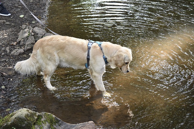
| Golden Retriever | Brain | Hemangiosarcoma | Lymphoma | Mast Cell Tumor | Melanoma | Oral | Osteosarcoma | Perianal/Anal Sac | Soft Tissue Sarcoma | |||
| Labrador Retriever | Hemangiosarcoma | Lymphoma | Mast Cell Tumor | Melanoma | Nasal Tumor | Oral | ||||||
| Scottish Terrier | Brain | Lymphoma | Melanoma | Nasal Tumor | Oral | Soft Tissue Sarcoma | TCC or UC | |||||
| Boxer | Brain (Glioma) | Hemangiosarcoma | Lymphoma | Mammary Tumor | Mast Cell Tumor | Oral | Osteosarcoma | Soft Tissue Sarcoma | ||||
| Beagle | Hemangiosarcoma | Lymphoma | Mast Cell Tumor | Perianal/Anal Sac | TCC or UC | |||||||
| West Highland White Terrier | Lymphoma | |||||||||||
| Chow Chow | Lymphoma | Melanoma | Oral | |||||||||
| Poodle, Standard | Lymphoma | Melanoma | Nasal Tumor | Oral | ||||||||
| Rottweiler | Lymphoma | Oral | Osteosarcoma | |||||||||
| Poodle, Toy | Lymphoma | Mammary Tumor | Melanoma | Nasal Tumor | ||||||||
| Yorkshire Terrier | Lymphoma | Mammary Tumor | ||||||||||
| German Shepherd | Hemangiosarcoma | Lymphoma | Mammary Tumor | Melanoma | Nasal Tumor | Oral | Osteosarcoma | Perianal/Anal Sac | ||||
| Poodle, Miniature | Lymphoma | Mammary Tumor | Melanoma | Nasal Tumor | Oral | |||||||
| Affenpinscher | ||||||||||||
| Afghan Hound | ||||||||||||
| Alaskan Malamute | Perianal/Anal Sac | |||||||||||
| American Eskimo, Toy and Standard | ||||||||||||
| American Foxhound | ||||||||||||
| American Pitt Bull Terrier | Hemangiosarcoma | |||||||||||
| American Staffordshire Terrier | ||||||||||||
| American Water Spaniel | ||||||||||||
| Anatolian Shepherd Dog | ||||||||||||
| Australian Cattle Dog | TCC or UC | |||||||||||
| Australian Shepherd | TCC or UC | |||||||||||
| Australian Terrier | ||||||||||||
| Basenji | ||||||||||||
| Bearded Collie | ||||||||||||
| Beauceron | ||||||||||||
| Bedlington Terrier | ||||||||||||
| Belgian Groenendael | ||||||||||||
| Belgian Malinois | ||||||||||||
| Belgian Tervuren | ||||||||||||
| Bernese Mountain Dog | Hemangiosarcoma | Melanoma | Soft Tissue Sarcoma | |||||||||
| Bichon Frise’ | TCC or UC | |||||||||||
| Black and Tan Coonhound | ||||||||||||
| Black Russian Terrier | ||||||||||||
| Bloodhound | Soft Tissue Sarcoma | |||||||||||
| Boerboel | ||||||||||||
| Border Collie | Brain | TCC or UC | ||||||||||
| Border Terrier | ||||||||||||
| Borzoi | Osteosarcoma | |||||||||||
| Boston Terrier | Brain | Mast Cell Tumor | Melanoma | Soft Tissue Sarcoma | ||||||||
| Bouvier des Flandres | Soft Tissue Sarcoma | |||||||||||
| Briard | ||||||||||||
| Brussels Griffon | ||||||||||||
| Bull Terrier | Mast Cell Tumor | Melanoma | ||||||||||
| Bull Terrier, Miniature | Mast Cell Tumor | Melanoma | ||||||||||
| Cairn Terrier | ||||||||||||
| Canaan Dog | ||||||||||||
| Cane Corso (Italian Mastiff) | ||||||||||||
| Caucasian Shepherd | ||||||||||||
| Cavalier King Charles Spaniel | Perianal/Anal Sac | |||||||||||
| Chesapeake Bay Retriever | Melanoma | |||||||||||
| Chinese Crested | ||||||||||||
| Chinese Shar-Pei | Mast Cell Tumor | Soft Tissue Sarcoma | TCC or UC | |||||||||
| Clumber Spaniel | ||||||||||||
| Curly Coated Retriever | ||||||||||||
| Dalmation | Hemangiosarcoma | |||||||||||
| Dandie Dinmont Terrier | ||||||||||||
| Dogo Argentino | ||||||||||||
| Dogue de Bordeaux | ||||||||||||
| English Foxhound | ||||||||||||
| English Toy Spaniel AKA King Charles Spaniel | ||||||||||||
| Field Spaniel | ||||||||||||
| Finnish Spitz | ||||||||||||
| Flat-Coated Retriever | Hemangiosarcoma | |||||||||||
| Fox Terrier, Smooth | Mast Cell Tumor | |||||||||||
| Fox Terrier, Toy | Mast Cell Tumor | |||||||||||
| Fox Terrier, Wire | TCC or UC | |||||||||||
| French Bulldog | ||||||||||||
| German Pinscher | ||||||||||||
| German Wirehaired Pointer | ||||||||||||
| Glen of Imaal Terrier | ||||||||||||
| Great Dane | Brain | Osteosarcoma | Soft Tissue Sarcoma | |||||||||
| Great Pyrenees | ||||||||||||
| Greater Swiss Mountain Dog | ||||||||||||
| Greyhound | Brain (Meningioma) | Hemangiosarcoma | Osteosarcoma | Soft Tissue Sarcoma | ||||||||
| Harrier | ||||||||||||
| Havanese | ||||||||||||
| Ibizan Hound | ||||||||||||
| Irish Terrier | ||||||||||||
| Irish Water Spaniel | ||||||||||||
| Irish Wolfhound | Osteosarcoma | |||||||||||
| Italian Greyhound | Brain | Hemangiosarcoma | ||||||||||
| Japanese Chin | ||||||||||||
| Keeshond | Nasal Tumor | |||||||||||
| Kerry Blue Terrier | ||||||||||||
| Komondor | ||||||||||||
| Kuvasz | ||||||||||||
| Lakeland Terrier | ||||||||||||
| Leonberger | Osteosarcoma | |||||||||||
| Lhasa Apso | TCC or UC | |||||||||||
| Lowchen | ||||||||||||
| Manchester Terrier Toy | ||||||||||||
| Manchester Terrier, Standard | ||||||||||||
| Mastiff | Brain | |||||||||||
| Miniature Pincher | ||||||||||||
| Neapolitan Mastiff | ||||||||||||
| Newfoundland | ||||||||||||
| Norfolk Terrier | ||||||||||||
| Norwegian Buhund | ||||||||||||
| Norwegian Elkhound | Brain | Nasal Tumor | ||||||||||
| Norwich Terrier | ||||||||||||
| Nova Scotia Duck Tolling Retriever | ||||||||||||
| Old English Sheepdog | Brain | |||||||||||
| Otterhound | ||||||||||||
| Papillon | ||||||||||||
| Parsons Russell Terrier | TCC or UC | |||||||||||
| Pekingese | Brain | |||||||||||
| Petit Basset Griffon Vendeen (PBGV) | ||||||||||||
| Pharaoh Hound | ||||||||||||
| Plott Hound | ||||||||||||
| Polish Lowland Sheepdog | ||||||||||||
| Pomeranian | ||||||||||||
| Portuguese Water Dog | Brain | Hemangiosarcoma | ||||||||||
| Presa Canario | ||||||||||||
| Pug | Brain | Mast Cell Tumor | Soft Tissue Sarcoma | |||||||||
| Puli | ||||||||||||
| Pyrenean Shepherd | ||||||||||||
| Rhodesian Ridgeback | Mast Cell Tumor | Soft Tissue Sarcoma | ||||||||||
| Saluki | ||||||||||||
| Samoyed | Perianal/Anal Sac | |||||||||||
| Schipperke | ||||||||||||
| Schnauzer, Miniature | Mast Cell Tumor | Melanoma | Perianal/Anal Sac | Soft Tissue Sarcoma | ||||||||
| Schnauzer, Standard | Mast Cell Tumor | Melanoma | Soft Tissue Sarcoma | |||||||||
| Sealyham Terrier | ||||||||||||
| Shiba Inu | ||||||||||||
| Shih Tzu | Brain | |||||||||||
| Siberian Husky | Perianal/Anal Sac | |||||||||||
| Silky Terrier | ||||||||||||
| Skye Terrier | Hemangiosarcoma | |||||||||||
| Soft-Coated Wheaten Terrier | ||||||||||||
| Spinone Italiano | ||||||||||||
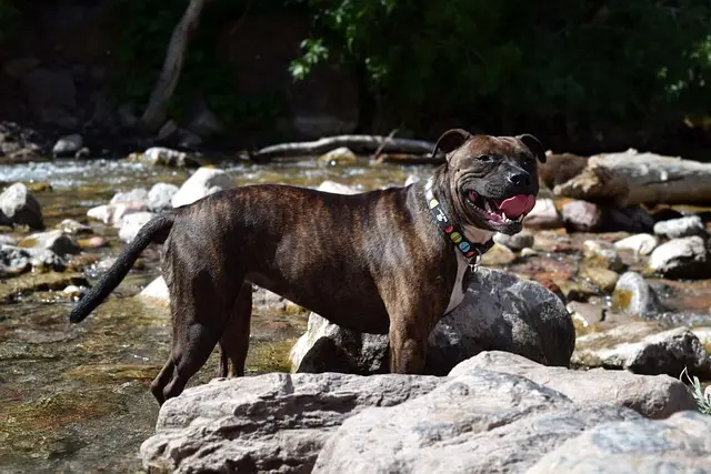
| Staffordshire Bull Terrier | Mast Cell Tumor | |||||||||||
| Sussex Spaniel | ||||||||||||
| Swedish Vallhund | ||||||||||||
| Tibetan Mastiff | ||||||||||||
| Tibetan Spaniel | ||||||||||||
| Tibetan Terrier | ||||||||||||
| Tosa | ||||||||||||
| Vizsla | Melanoma | |||||||||||
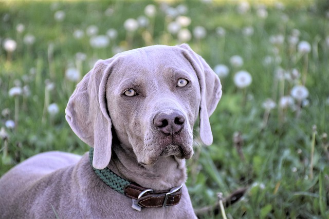
| Weimaraner | Mast Cell Tumor | |||||||||||
| Welsh Corgi, Cardigan | ||||||||||||
| Welsh Corgi, Pembroke | ||||||||||||
| Welsh Springer Spaniel | ||||||||||||
| Welsh Terrier | ||||||||||||
| Whippet | Hemangiosarcoma | TCC or UC | ||||||||||
| Wirehaired Pointing Griffon | ||||||||||||
| Akita (American) | Oral | |||||||||||
| Collie, Rough / Smooth Coat | Brain (Meningioma) | Nasal Tumor | Oral | TCC or UC | ||||||||
| Gordon Setter | Melanoma | Oral | ||||||||||
| Irish Setter | Melanoma | Oral | Osteosarcoma | Soft Tissue Sarcoma | ||||||||
| Schnauzer, Giant | Mast Cell Tumor | Melanoma | Oral | |||||||||
| Scottish Deerhound | Brain | Melanoma | Oral | Osteosarcoma | TCC or UC | |||||||
| Shetland Sheepdog | Nasal Tumor | Oral | TCC or UC | |||||||||
| Brittany | Mammary Tumor | |||||||||||
| Chihuahua | Mammary Tumor | Melanoma | ||||||||||
| English Cocker Spaniel | Mammary Tumor | |||||||||||
| English Setter | Mammary Tumor | |||||||||||
| English Springer Spaniel | Mammary Tumor | Melanoma | Perianal/Anal Sac | |||||||||
| Maltese | Mammary Tumor | |||||||||||
| Pointer | Hemangiosarcoma | Mammary Tumor | ||||||||||
| Cocker Spaniel (American) | Mammary Tumor | Mast Cell Tumor | Melanoma | Oral | Perianal/Anal Sac | |||||||
| Dachshund | Brain | Mammary Tumor | Mast Cell Tumor | Oral | Perianal/Anal Sac | |||||||
| Doberman Pinscher | Brain | Mammary Tumor | Melanoma | Oral | Osteosarcoma | |||||||
| German Shorthaired Pointer | Mammary Tumor | Nasal Tumor | Oral |