Canine Pulmonic Stenosis
& Breeds at Risk
Research, Resources & Education
This website is based on research and is NOT created to diagnose your pet.
Each animal is an individual and may exhibit symptoms in a different way.
It is advised that you ALWAYS CHECK WITH YOUR VETERINARIAN for a proper diagnosis and treatment plan.
Table of Contents
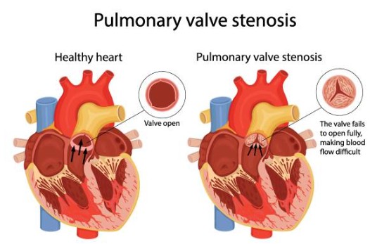
Pulmonic Stenosis (PS)
Pulmonic stenosis is a common congenital defect of dogs, and it most commonly involves fusion or dysplasia of the pulmonic valve leaflets (valvar or valvular).
Many dogs have no clinical signs. Dogs with mild disease never develop any problems and may live a normal lifespan. However, dogs with advanced disease may have exercise intolerance, collapsing, arrhythmias, or heart failure.
Treatment: Animals with moderate or severe pulmonic stenosis can benefit from balloon valvuloplasty or surgical intervention and/or can be controlled with medications
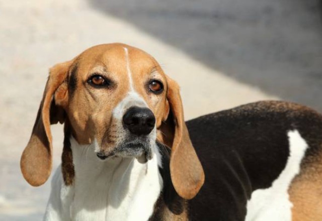
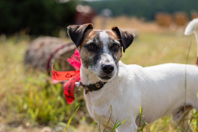
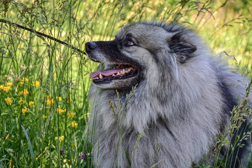
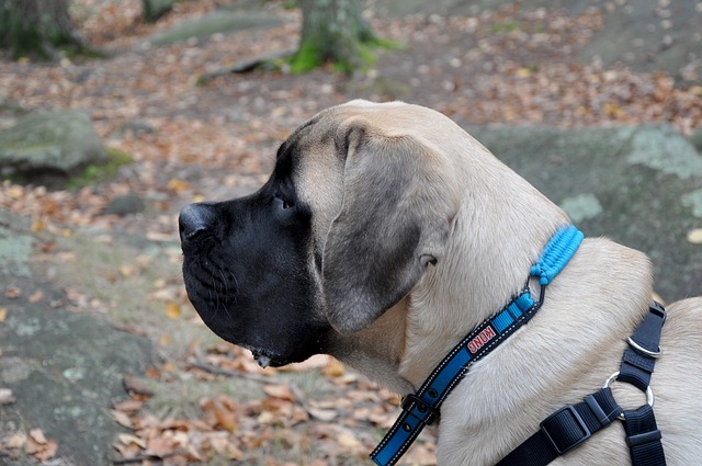
What Dogs are at Risk?
Some Dogs that are at Risk due to Genetic Predisposition


What is Pulmonic Stenosis?
Pulmonic stenosis (Merck Professional)
Pulmonic stenosis – Merck Professional
Pulmonic stenosis is a common congenital defect of dogs, and it most commonly involves fusion or dysplasia of the pulmonic valve leaflets (valvar or valvular).
- This condition affects a variety of dog breeds and is typically associated with a loud ejection murmur heard over the left base.
- Impedance to normal ejection and pressure overload of the right ventricle can result in right-side CHF, low-output signs (exercise intolerance, exertional syncope), and cardiac arrhythmias.
- Diagnosis can be made by echocardiography. Prognosis varies with severity of disease, and balloon valvuloplasty is typically recommended for moderate to severe cases.
- Pulmonic stenosis is common in dogs and infrequent in cats.
- It results in obstruction to right ventricular outflow due, in most cases, to commissural fusion or dysplasia of the pulmonic valve with or without annular hypoplasia.
- Although the valvular form is most common, stenosis can also occur in the subvalvular region (infundibulum) or in the supravalvular area.
There are two main types of PS: (Improve Veterinary Practice)
There are two main types of PS: – Improve Veterinary Practice
- Valvular – affecting the leaflets that are fused together
- Mixed – can show a valvular component with a narrowing that can be due to a hypoplastic pulmonic annulus or narrowing above or below the valve
- In some breeds, such as the English Bulldog, the stenosis can be worsened by the presence of an abnormal coronary artery that wraps around the pulmonary artery (Buchanan, 1990).
- The severity of pulmonary stenosis is classified by the pressure gradient (PG) across the valve, which is estimated using the Bernoulli equation
Pulmonic Stenosis Videos
YouTube Videos that help explain Pulmonic Stenosis (PS) in Dogs
Disclaimer:
This is for research only and Lost Temple Pets does not endorse any video presented on this website.
It is advised that you ALWAYS CHECK WITH YOUR VETERINARIAN for a proper diagnosis and treatment plan.
Playlist
Causes
Causes (Cornell)
Causes Cornell
Pulmonic stenosis is a congenital heart defect of the semilunar valve that is between the right ventricle and the pulmonary artery (great vessel that takes blood to the lungs).
- The leaflets of this valve are thickened and/or partially fused together. Sometimes the supporting structure known as the annulus is also narrow.
- Dogs that have this congenital defect have a wide range of stenosis to include very mild to severe obstruction to blood flow from the heart to the lungs.
- This defect may be associated with other congenital defects too (e.g. ventricular septal defect, overriding aorta, subaortic stenosis).
- Because this disease is associated with certain breeds it is likely that it is at least in part due to a mutation in as yet unidentified gene.
Symptoms / Signs
Clinical Findings (Merck Veterinary Manual (Professional)
Clinical Findings – Merck Veterinary Manual (Professional)
Animals with pulmonic stenosis may have a history of exercise intolerance and failure to thrive.
- Right-side CHF may be present and is characterized by ascites and less commonly peripheral edema.
- A prominent systolic ejection murmur is present and heard loudest over the pulmonic valve area (left base).
- A corresponding precordial thrill may be present. Jugular distention and pulsations may also be present.
Clinical Signs (Cornell)
Clinical Signs – Cornell
- Many dogs have no clinical signs. Dogs with mild disease never develop any problems and may live a normal lifespan.
- However, dogs with advanced disease may have exercise intolerance, collapsing, arrhythmias, or heart failure.
Diagnosis
Diagnostic evaluation (Improve Veterinary Practice)
Diagnostic evaluation – Improve Veterinary Practice
- Echocardiography is the best diagnostic tool to diagnose PS as it allows practitioners to evaluate the severity of the stenosis by measuring velocities across the valve, as well as any secondary changes such as right ventricular hypertrophy.
- Concomitant congenital diseases, such as tricuspid dysplasia, are possible and should be taken into consideration when creating future plans.
- Electrocardiography (ECG) might be required to investigate ventricular arrhythmias and/or atrial fibrillation, which can be associated with PS – especially in severe cases. A deep S wave on ECG is also a common finding in dogs with right ventricular hypertrophy.
Diagnosis (Cornell)
Diagnosis – Cornell
Virtually all dogs with clinically important pulmonic stenosis will have a cardiac murmur heard when the chest is listened to with a stethoscope. This is auscultation of the chest.
- Often, but not always, how loud the murmur is in this particular disease correlates with severity.
- An important example of the exception to this general statement is with tetralogy of Fallot where several congenital defects are present together with pulmonic stenosis.
- Radiography and angiocardiography.
- Radiographs which are made with x-rays provide information regarding the size and shape of the silhouette of the heart.
- Angiocardiography is a type of radiography whereby contrast (dye) is injected into the vasculature to see the stenosis. This is most often done at the time of treatment using balloon valvuloplasty.
- Echocardiography An important diagnostic tool to fully characterize the structure and function of the pulmonic valve and the support structures involves the ultrasound of the heart known as echocardiography.
- This test permits the examination not only of the muscle and valve, but also of the blood flow (Doppler echocardiography). Determination of the blood flow across the stenotic valve is important to learn the severity of the pulmonic stenosis because this guides the recommendations for treatment.
- Electrocardiography An electrocardiogram (ECG) may be preformed to further characterize dogs with pulmonic stenosis; however, this test is usually not as important as the physical examination, radiograph, and echocardiogram.
Treatment
Treatment of Pulmonic Stenosis in Animals (Merck Veterinary Manual (Professional)
Treatment of Pulmonic Stenosis in Animals – Merck Veterinary Manual (Professional)
Moderate or severe cases can be treated with balloon valvuloplasty or surgery.
- Animals with moderate or severe pulmonic stenosis can benefit from balloon valvuloplasty or surgical intervention (valvulotomy, patch grafting, partial valvulectomy, or conduits).
- Balloon valvuloplasty is a minimally invasive and highly effective treatment option.
- Some animals are medically managed with a beta-blocker (eg, atenolol) alone or in conjunction with balloon valvuloplasty.
- Congestive heart failure should be medically managed if present.
- Similarly, the presence of supraventricular or ventricular arrhythmias warrants therapy with the appropriate antiarrhythmic drug(s).
Medical or surgical therapy (Improve Veterinary Practice)
Medical or surgical therapy – Improve Veterinary Practice
- Medical management aims to alleviate clinical signs (ie collapse, exercise intolerance) and improve quality of life.
- Beta-adrenergic blockers, such as atenolol, are commonly prescribed to reduce heart rate and myocardial oxygen demand. These must be gradually introduced and never stopped suddenly.
- Diuretics may be indicated for managing signs of right-sided heart failure.
- However, medical therapy alone is often insufficient for addressing severe cases of PS, and the disease tends to progress without a surgical resolution.
- Surgical intervention is considered the treatment of choice for moderate to severe PS. Balloon valvuloplasty (BVP), a minimally invasive procedure, involves the insertion of a balloon catheter into the stenotic pulmonary valve and inflation to dilate the narrowed area.
- This technique has demonstrated excellent short- and long-term outcomes in dogs with valvular stenosis, with significant improvements in clinical signs and survival rates.
- Complications of BVP include arrhythmias and haemorrhage.
- Moreover, some dogs might not fully respond to BVP with only a minor reduction in PS severity (reduction of 50 percent or more is considered successful).
- In cases where BVP is unlikely to be successful (such as in cases with mixed-type PS), pulmonary artery stenting may be pursued.
- This is a minimally invasive technique that involves the implantation of a metal stent across the stenotic lesion (Borgeat et al., 2021).
- Complications for this procedure, besides those listed for BVP, include stent fracture, migration and crimping.
- Open-heart surgery for right ventricular outflow tract grafting with an expanded polytetrafluoroethylene patch under cardiopulmonary bypass has been described as well (Bristow et al., 2018).
References
References
animalia-life.club – Picture
https://animalia-life.club/qa/pictures/how-long-will-my-dog-live-with-pulmonic-stenosis
Cornell College of Veterinary Medicine – Pulmonic Stenosis in Dogs
https://www.vet.cornell.edu/hospitals/services/cardiology/pulmonic-stenosis-dogs
Improve Veterinary Practice – Congenital cardiac defects in dogs: pulmonic stenosis and atrial septal defects
by Mattia Basili
29 April 2024
Merck Veterinary Manual (Pet Owner Version) – Congenital and Inherited Disorders of the Cardiovascular System in Dogs
By Sandra P. Tou, DVM, DACVIM-Cardiology, DACVIM-SAIM, Department of Clinical Sciences, College of Veterinary Medicine, North Carolina State University
Reviewed/Revised Jun 2018
Merck Veterinary Manual (Professional Version) – Pulmonic Stenosis in Animals
By Sandra P. Tou, DVM, DACVIM-Cardiology, DACVIM-SAIM
Reviewed/Revised Jan 2020
Veterian Key –Pulmonic Stenosis
Amara H. Estrada, Gainesville, Florida
Herbert W. Maisenbacher, III, Gainesville, Florida
https://veteriankey.com/chapter-65-pulmonic-stenosis/
VIDEOS
Dr. Bozelka, ER Veterinarian – Pulmonic Stenosis in Pets
https://www.youtube.com/watch?v=qf9nhLbACXY
KK Women’s and Children’s Hospital – Heart Conditions – Pulmonary Stenosis
https://www.youtube.com/watch?v=Rjorbxccxt0&t=7s
mikemartinheartvet – Balloon catheter dilatation of pulmonic stenosis in a dog
Cardiac/Breed Chart
| BREED | Atrial septal defect (ASD) | Chronic mitral valvular disease (CMVDz) | Dilated cardiomyopathy (DCM) | Mitral valvular dysplasia (MVD) | Patent ductus arteriosus (PDA) | Pulmonic stenosis (PS) | Subaortic stenosis (SAS) | Tricuspid valvular dysplasia (TVD) | |
|---|---|---|---|---|---|---|---|---|---|
| Affenpinscher | Patent ductus arteriosus (PDA) | ||||||||
| Afghan Hound | Dilated cardiomyopathy (DCM) | Mitral valvular dysplasia (MVD) | |||||||
| Airedale Terrier | |||||||||
| Akita (American) | |||||||||
| Alaskan Malamute | |||||||||
| American Eskimo, Toy and Standard | |||||||||
| American Foxhound | |||||||||
| American Pitt Bull Terrier | |||||||||
| American Staffordshire Terrier | |||||||||
| American Water Spaniel | |||||||||
| Anatolian Shepherd Dog | |||||||||
| Australian Cattle Dog | |||||||||
| Australian Shepherd | |||||||||
| Australian Terrier | |||||||||
| Basenji | |||||||||
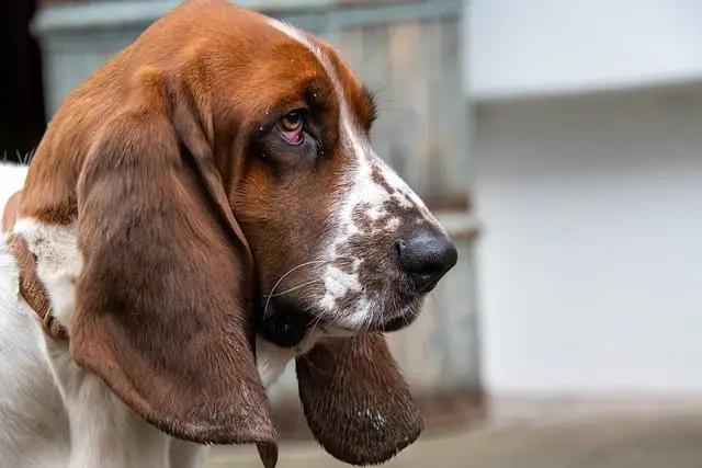
| Basset Hound | Pulmonic stenosis (PS) | ||||||||
| Beagle | Pulmonic stenosis (PS) | ||||||||
| Bearded Collie | |||||||||
| Beauceron | |||||||||
| Bedlington Terrier | |||||||||
| Belgian Groenendael | |||||||||
| Belgian Malinois | |||||||||
| Belgian Tervuren | |||||||||
| Bernese Mountain Dog | |||||||||
| Bichon Frise’ | Patent ductus arteriosus (PDA) | ||||||||
| Black and Tan Coonhound | |||||||||
| Black Russian Terrier | |||||||||
| Bloodhound | |||||||||
| Boerboel | |||||||||
| Border Collie | |||||||||
| Border Terrier | |||||||||
| Borzoi | |||||||||
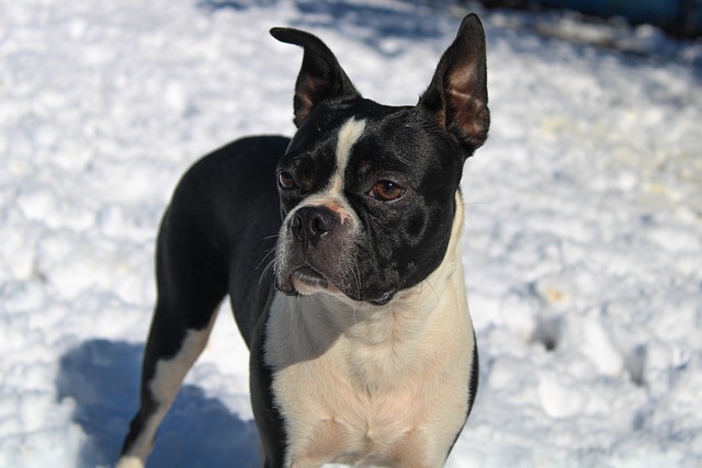
| Boston Terrier | Pulmonic stenosis (PS) | ||||||||
| Bouvier des Flandres | Subaortic stenosis (SAS) | ||||||||
| Boxer | Atrial septal defect (ASD) | Dilated cardiomyopathy (DCM) | Pulmonic stenosis (PS) | Subaortic stenosis (SAS) | |||||
| Briard | |||||||||
| Brittany | |||||||||
| Brussels Griffon | |||||||||
| Bull Terrier | Mitral valvular dysplasia (MVD) | ||||||||
| Bull Terrier, Miniature | |||||||||
| Bulldog, English | Pulmonic stenosis (PS) | Subaortic stenosis (SAS) | |||||||
| Bullmastiff | |||||||||
| Cairn Terrier | |||||||||
| Canaan Dog | |||||||||
| Cane Corso (Italian Mastiff) | |||||||||
| Caucasian Shepherd | |||||||||
| Cavalier King Charles Spaniel | Chronic mitral valvular disease (CMVDz) | Patent ductus arteriosus (PDA) | |||||||
| Chesapeake Bay Retriever | |||||||||
| Chihuahua | Chronic mitral valvular disease (CMVDz) | Patent ductus arteriosus (PDA) | Pulmonic stenosis (PS) | ||||||
| Chinese Crested | |||||||||
| Chinese Shar-Pei | |||||||||
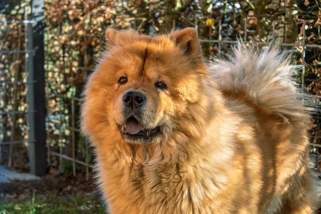
| Chow Chow | Pulmonic stenosis (PS) | ||||||||
| Clumber Spaniel | |||||||||
| Cocker Spaniel (American) | Chronic mitral valvular disease (CMVDz) | Dilated cardiomyopathy (DCM) | Patent ductus arteriosus (PDA) | Pulmonic stenosis (PS) | |||||
| Collie, Rough / Smooth Coat | Patent ductus arteriosus (PDA) | ||||||||
| Curly Coated Retriever | |||||||||
| Dachshund | Chronic mitral valvular disease (CMVDz) | ||||||||
| Dalmation | |||||||||
| Dandie Dinmont Terrier | |||||||||
| Doberman Pinscher | Atrial septal defect (ASD) | Dilated cardiomyopathy (DCM) | |||||||
| Dogo Argentino | |||||||||
| Dogue de Bordeaux (Mastiff) | |||||||||
| English Cocker Spaniel | Dilated cardiomyopathy (DCM) | Pulmonic stenosis (PS) | |||||||
| English Foxhound | |||||||||
| English Setter | |||||||||
| English Springer Spaniel | Dilated cardiomyopathy (DCM) | Patent ductus arteriosus (PDA) | |||||||
| English Toy Spaniel AKA King Charles Spaniel | |||||||||
| Field Spaniel | |||||||||
| Finnish Spitz | |||||||||
| Flat-Coated Retriever | |||||||||
| Fox Terrier, Smooth | |||||||||
| Fox Terrier, Toy | |||||||||
| Fox Terrier, Wire | Pulmonic stenosis (PS) | ||||||||
| French Bulldog | |||||||||
| German Pinscher | |||||||||
| German Shepherd | Dilated cardiomyopathy (DCM) | Mitral valvular dysplasia (MVD) | Patent ductus arteriosus (PDA) | Subaortic stenosis (SAS) | Tricuspid valvular dysplasia (TVD) | ||||
| German Shorthaired Pointer | Subaortic stenosis (SAS) | ||||||||
| German Wirehaired Pointer | |||||||||
| Glen of Imaal Terrier | |||||||||
| Golden Retriever | Dilated cardiomyopathy (DCM) | Subaortic stenosis (SAS) | |||||||
| Gordon Setter | |||||||||
| Great Dane | Dilated cardiomyopathy (DCM) | Mitral valvular dysplasia (MVD) | Subaortic stenosis (SAS) | Tricuspid valvular dysplasia (TVD) | |||||
| Great Pyrenees | |||||||||
| Greater Swiss Mountain Dog | |||||||||
| Greyhound | |||||||||
| Harrier | |||||||||
| Havanese | |||||||||
| Ibizan Hound | |||||||||
| Irish Setter | Tricuspid valvular dysplasia (TVD) | ||||||||
| Irish Terrier | Patent ductus arteriosus (PDA) | ||||||||
| Irish Water Spaniel | |||||||||
| Irish Wolfhound | Dilated cardiomyopathy (DCM) | ||||||||
| Italian Greyhound | |||||||||
| Japanese Chin | |||||||||
| Keeshond | Patent ductus arteriosus (PDA) | Pulmonic stenosis (PS) | |||||||
| Kerry Blue Terrier | Patent ductus arteriosus (PDA) | ||||||||
| Komondor | |||||||||
| Kuvasz | |||||||||
| Labrador Retriever | Dilated cardiomyopathy (DCM) | Patent ductus arteriosus (PDA) | Pulmonic stenosis (PS) | Tricuspid valvular dysplasia (TVD) | |||||
| Lakeland Terrier | |||||||||
| Lhasa Apso | Chronic mitral valvular disease (CMVDz) | ||||||||
| Lowchen | |||||||||
| Maltese | Chronic mitral valvular disease (CMVDz) | Patent ductus arteriosus (PDA) | |||||||
| Manchester Terrier Toy | |||||||||
| Manchester Terrier, Standard | |||||||||
| Mastiff, English | Dilated cardiomyopathy (DCM) | Pulmonic stenosis (PS) | |||||||
| Miniature Pincher | |||||||||
| Neapolitan Mastiff | |||||||||
| Newfoundland | Dilated cardiomyopathy (DCM) | Patent ductus arteriosus (PDA) | Pulmonic stenosis (PS) | Subaortic stenosis (SAS) | |||||
| Norfolk Terrier | |||||||||
| Norwegian Buhund | |||||||||
| Norwegian Elkhound | |||||||||
| Norwich Terrier | |||||||||
| Nova Scotia Duck Tolling Retriever | |||||||||
| Old English Sheepdog | Atrial septal defect (ASD) | Dilated cardiomyopathy (DCM) | Tricuspid valvular dysplasia (TVD) | ||||||
| Otterhound | |||||||||
| Papillon | Chronic mitral valvular disease (CMVDz) | ||||||||
| Parsons Russell Terrier | |||||||||
| Pekingese | Chronic mitral valvular disease (CMVDz) | ||||||||
| Petit Basset Griffon Vendeen (PBGV) | |||||||||
| Pharaoh Hound | |||||||||
| Plott Hound | |||||||||
| Pointer | Subaortic stenosis (SAS) | ||||||||
| Polish Lowland Sheepdog | |||||||||
| Pomeranian | Chronic mitral valvular disease (CMVDz) | Patent ductus arteriosus (PDA) | |||||||
| Poodle, Miniature | Chronic mitral valvular disease (CMVDz) | Patent ductus arteriosus (PDA) | |||||||
| Poodle, Standard | Atrial septal defect (ASD) | Patent ductus arteriosus (PDA) | |||||||
| Poodle, Toy | Chronic mitral valvular disease (CMVDz) | Patent ductus arteriosus (PDA) | |||||||
| Portuguese Water Dog | Dilated cardiomyopathy (DCM) | ||||||||
| Presa Canario | |||||||||
| Pug | |||||||||
| Puli | |||||||||
| Pyrenean Shepherd | |||||||||
| Rhodesian Ridgeback | |||||||||
| Rottweiler | Subaortic stenosis (SAS) | ||||||||
| Saluki | |||||||||
| Samoyed | Atrial septal defect (ASD) | Pulmonic stenosis (PS) | Subaortic stenosis (SAS) | ||||||
| Schipperke | |||||||||
| Schnauzer, Miniature | Chronic mitral valvular disease (CMVDz) | Pulmonic stenosis (PS) | |||||||
| Schnauzer, Giant | Pulmonic stenosis (PS) | ||||||||
| Schnauzer, Standard | |||||||||
| Scottish Deerhound | Dilated cardiomyopathy (DCM) | ||||||||
| Scottish Terrier | |||||||||
| Sealyham Terrier | |||||||||
| Shetland Sheepdog | Patent ductus arteriosus (PDA) | ||||||||
| Shiba Inu | |||||||||
| Shih Tzu | Chronic mitral valvular disease (CMVDz) | ||||||||
| Siberian Husky | |||||||||
| Silky Terrier | |||||||||
| Skye Terrier | |||||||||
| Soft-Coated Wheaten Terrier | |||||||||
| Spinone Italiano | |||||||||
| St. Bernard | Dilated cardiomyopathy (DCM) | ||||||||
| Staffordshire Bull Terrier | |||||||||
| Sussex Spaniel | |||||||||
| Swedish Vallhund | |||||||||
| Tibetan Mastiff | |||||||||
| Tibetan Spaniel | |||||||||
| Tibetan Terrier | |||||||||
| Tosa | |||||||||
| Vizsla | |||||||||
| Weimaraner | |||||||||
| Welsh Corgi, Cardigan | Patent ductus arteriosus (PDA) | ||||||||
| Welsh Corgi, Pembroke | Patent ductus arteriosus (PDA) | ||||||||
| Welsh Springer Spaniel | |||||||||
| Welsh Terrier | |||||||||
| West Highland White Terrier | Chronic mitral valvular disease (CMVDz) | Pulmonic stenosis (PS) | |||||||
| Whippet | |||||||||
| Wirehaired Pointing Griffon | |||||||||
| Yorkshire Terrier | Chronic mitral valvular disease (CMVDz) | Patent ductus arteriosus (PDA) | |||||||
| Spanish Mastiff | |||||||||
| Treeing Walker Coonhound | |||||||||
| Barbet | |||||||||
| Cirneco dell'Etna | |||||||||
| Broholmer | |||||||||
| Leonberger | |||||||||
| Rat Terrier | |||||||||
| Xoloitzcuintli | |||||||||
| Dutch Shepherd |
This consists of abnormal tissue located just below the aortic valve that creates an obstruction the heart has to overcome to pump blood out to the body.