
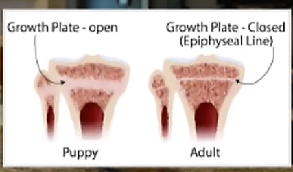
Growth Plates
Growth plates in pets are crucial for bone development and are influenced by hormones. They allow for the lengthening of bones as the pet grows. Once the animal reaches maturity, these plates ossify and close, marking the end of bone growth.
What are Growth Plates?
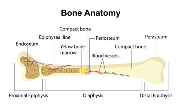
- Location: Growth plates, or epiphyseal plates, are areas of cartilage located at the ends of long bones , such as the femur, tibia, and humerus in young animals.
- Function: They consist of cartilage cells that actively divide, allowing for the lengthening of bones as the dog grows. This process is essential during the puppy and young dog stages of development.
Hormonal Influence
Hormones play a significant role in regulating the growth and closure of these plates:
- Growth Hormone (GH): Produced by the pituitary gland, GH stimulates growth and development. An excess of GH can lead to abnormal growth patterns.
- Thyroid Hormones: These hormones are essential for normal growth and metabolism. Imbalances can affect growth plate function.
- Sex Hormones: Estrogen and testosterone influence the timing of growth plate closure, which varies by breed and size.
Nutritional Factors
Proper nutrition is crucial during this growth phase to ensure healthy bone development. A balanced diet rich in calcium and phosphorus supports bone strength
Importance of Growth Plates
Importance of Growth Plates
Understanding growth plates is crucial for managing a dog’s exercise and weight-bearing activities to prevent injuries before they close.
- Many bones have multiple growth plates, so skeletal health depends on the uninterrupted closure of over 700 growth plates. These plates are told to stop developing by hormones contained in and released by the reproductive organs.
- Identical bones in both legs develop at the same time, however the various bones within each limb do not.
- Your puppy’s bones are not finished developing just because she has reached mature height. Bones will continue ‘filling in’ even after they have reached their desired length.
- Bones grow first and then muscles and connective tissue second. The time following a growth spurt is when puppy is most vulnerable for hyperextension and impact injuries as the muscles, tendons and ligaments are struggling to support bones which are now too long until they can catch up.
- Early neutering (which is any time before the last growth plates close @ 14-18 months of age) will disrupt balanced, healthy growth as these hormonal messages are removed along with the organs. (see below)
Growth Plate Closure and Injury Risks
Different bones and limb segments close at varying times, creating asymmetrical growth and imbalance.
- Joints including knees, hips, elbows, and wrists are vulnerable during their respective closure windows, especially around 6 months.
- Asymmetrical closure causes joint imbalance, increasing risks of dysplasia, sprains, and pelvic issues
- Damage to growth plates can lead to long-term issues, including deformities or uneven growth
Hormone Effects on Growth Plates
Hormone effects on growth plates
Hormones play a central role in regulating the growth and closure of growth plates in bones, especially during puppy development:
- Growth Hormone (GH): Produced by the pituitary gland, GH stimulates the division of cartilage cells in the growth plates, promoting bone lengthening and overall skeletal growth. Excess GH can cause abnormal growth patterns.
- Thyroid Hormones: Essential for normal growth and metabolism, these hormones help regulate the pace of growth plate activity. Imbalances can disrupt normal growth plate function, leading to stunted or excessive growth.
- Sex Hormones (Estrogen and Testosterone): These hormones, released by the reproductive organs, are responsible for signaling the closure of growth plates at the end of the growth period. The timing of growth plate closure varies by breed and size, but generally, as levels of estrogen and testosterone rise with maturity, growth plates ossify and bone lengthening stops.
Practical Implications
- Growth plates are highly sensitive to hormonal changes. Disruption of normal hormone levels (such as through early neutering) can lead to abnormal bone development and increased risk of skeletal injuries.
- Balanced hormone levels are crucial for the healthy and timely closure of growth plates, ensuring proper bone length and joint function. Understanding these hormonal effects is essential for managing growth, exercise, and medical decisions in growing puppies.
Risks of Early Neutering
Risks of Early Neutering
Early neutering (removal of reproductive organs before growth plates close, typically before 14–18 months) removes these hormonal signals, causing growth plates to remain open longer.
- Disrupted Bone Growth and Imbalance: Sex hormones (estrogen and testosterone) are responsible for signaling growth plates to close at the appropriate time. Early neutering removes these hormones, causing growth plates that have not yet closed to remain open longer than normal. This results in bones growing longer than intended, leading to disproportionate limb length and skeletal imbalance.
- Strained Connective Tissue: As bones continue to lengthen, muscles, tendons, and ligaments struggle to keep up, often becoming overstretched or strained. This can leave joints less stable and more prone to injury.
- Increased Risk of Joint Disorders: The imbalance between bone and soft tissue growth increases the risk of joint problems such as hip and elbow dysplasia, wrist sprains, and pelvic dysfunction. Joints like the knees, hips, hocks, and wrists are particularly vulnerable during this period.
- Prolonged Vulnerability to Injury: With growth plates closing at different times across various bones, puppies experience a prolonged window of vulnerability to injuries, especially during high-impact activities. This can lead to chronic compensation or re-injury patterns affecting other joints and the spine.
- Potential for Long-Term Deformities: Damage or disruption to growth plates during this extended growth phase can result in permanent deformities or uneven limb development.
Injury Risks During Growth
Injury Risks During Growth
- Bones, tendons, and ligaments develop at different rates, requiring careful management to prevent long-term damage.
- Imbalanced and asymmetrical growth increases vulnerability to injuries during active puppy stages with risk of dysplasia, sprains, and joint issues.
- Avoid high-impact activities before growth plates close to prevent injuries and long-term skeletal problems.
- Activities like jumping and stairs should be limited until bones fully mature.
- Early injury or trauma can disrupt proper formation, leading to deformities or chronic issues later.
Puppies should avoid high-impact and strenuous exercises to protect their developing joints and overall health.
Activities to Avoid
- Long Runs or Hikes: Puppies should not engage in prolonged running or hiking, as it can strain their joints.
- Jumping: Avoid activities that require jumping, such as agility training or jumping over or off obstacles, which can lead to injuries.
- Intense Fetch Games: While fetch can be fun, overly vigorous games can lead to exhaustion and joint stress.
- Heavy Weight-Bearing Exercises: Activities that involve carrying weights or excessive resistance should be avoided
Summary
- Proper nutrition, controlled activity, and awareness of growth plate timing are essential for healthy development.
- Proper management of exercise and nutrition is essential to prevent long-term skeletal issues.
Vulnerable Growth Plates
The knees, hips, hocks and wrists are particularly vulnerable by the time puppy is 6 months old as some bones have finished growing while others have a while longer to close.
- Avoid high impact activities including jumping off the furniture and stairs, as it increases the risk for micro-injuries which contribute to dysplasia in hips, elbows and shoulders, wrist sprains and pelvic dysfunction.
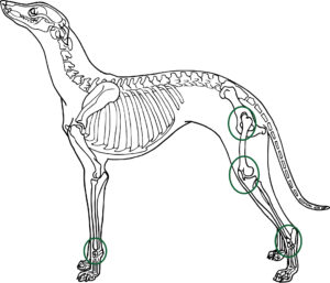
Growth Plates - Asymmetrical Closure Times
Asymmetrical growth plate closures can significantly impact skeletal development and final height.
- This phenomenon occurs when growth plates in different bones close at varying rates, and never at the same time or place, which can lead to discrepancies in limb length and overall body proportions.
- Because of the different gaps in closure times, you will find that the puppy’s skeleton will always be off balance until 14 – 18 months of age.
- Muscles, tendons and ligaments each also grow at their own rate and they respond differently to exercise stress
Consequences
- Limb Length Discrepancies: Asymmetrical closure can result in one limb being shorter than the other, which may require orthopedic intervention.
- Postural Issues: Uneven growth can lead to postural problems and affect overall biomechanics.
Causes of Asymmetrical Closure
- Genetic Factors: Genetic predispositions can influence the timing and pattern of growth plate closure.
- Hormonal Influences: Hormones such as growth hormone and sex hormones play crucial roles in regulating growth plate activity.
- Injuries: Trauma to a growth plate can lead to premature closure in the affected area, resulting in asymmetry.
- Medical Conditions: Certain conditions, like skeletal dysplasia’s or endocrine disorders, can also affect growth plate behavior.

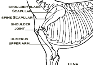
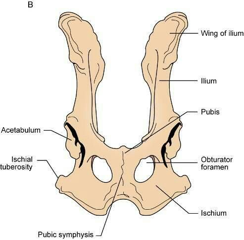
SHOULDER BLADE and PELVIS BONE (also see Pelvis below)
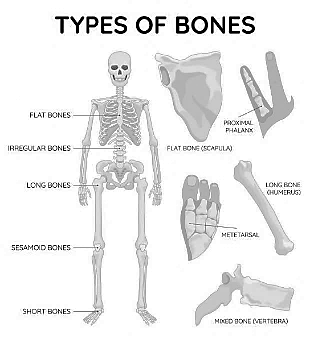
Flat, spongy bones (skull, shoulder blades, pelvis) have multiple growth plates closing at different times, with some extending into late age. These growth plates are not as strong as compact bone.
- Flat bones may have one big sheath of growth plate because they don’t just grow up and down but outwards and in depth as well.
- Shoulder blade closes between 3.9 and 7 months.
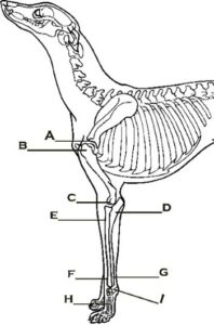
- Upper end of the humerus (B) closes between 9.1 and 15 months. (Lower end of the humerus closes between 4.6 and 7.8 months).
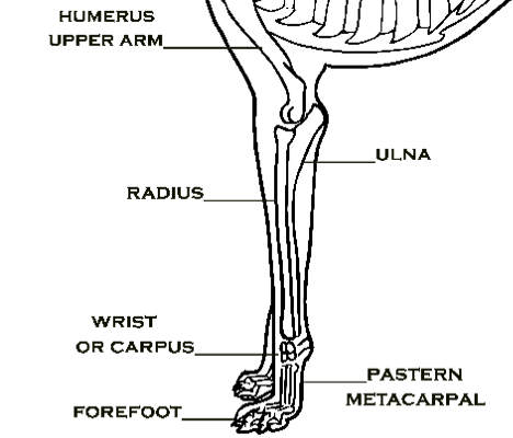
ELBOW - Front
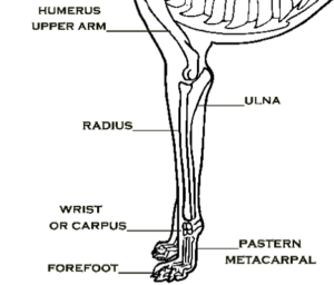
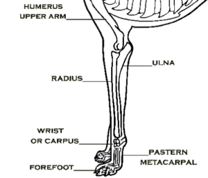
This joint is made up of three bones, the humerus, radius and ulna. They must work together to create smooth, fluid movements, while developing at very different times.
- Humerus (C), lower end of the long bone, closes between 4.6 and 11 months.
- Radius (E – top) closes between 4.5 and 11 months.
- Ulna (D – top) closes between 5.3 and 15 months.
- The elbow joint develops over a 4.5 – 15 month period, with bones maturing at different times, increasing injury risk.
- Asymmetrical bone development creates imbalance in the muscles, tendons and ligaments, which are not growing at the same rate as the bones.
- This increases the risk of injury going down the stairs, jumping off furniture, in and out of vehicles and jumping when chasing balls and other objects.
CARPUS / WRIST - Front
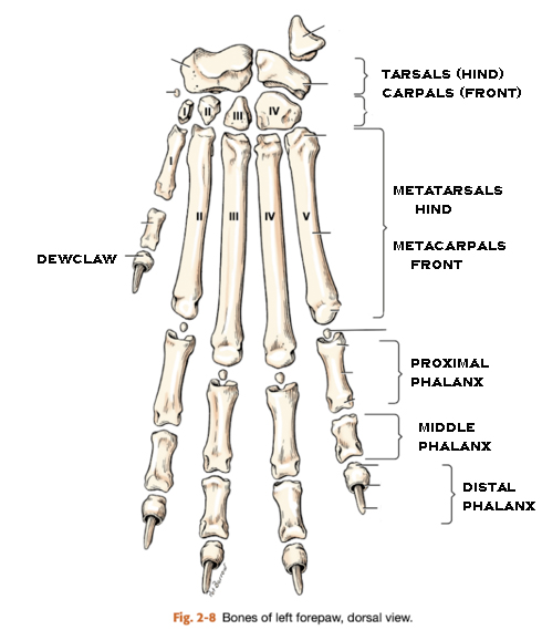
The wrist and paw bones close early around 3-9 months, but some continue growing up to 17 months, affecting stability.
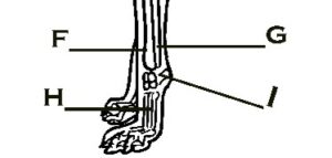
- Radial (carpal/bottom – F) closes between 4.5 and 17 months
- Ulna (carpal/bottom – G) closes between 7.2-15
- Wrist – Carpals (I) closes around 3.3 months
- Metacarpals (H) closes between 6.7 and 7 months.
- The wrist closes at around 3.3 months while the bones above it continue growing for up to another 14 months, to provide stability at the wrist during weight bearing.
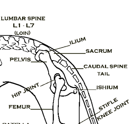
HIP - Hind / Back
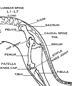
The canine hip joint is a ball-and-socket joint, where the femoral head (the ball) fits into the acetabulum (the socket) of the pelvis. This structure allows for a wide range of motion, which is crucial for a dog’s agility and activity levels.
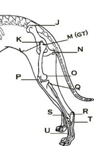
- Acetabulum (socket- K) that the head of the femur fits into closes between 3.7 and 6 months.
- Femoral Head (sits inside the socket – L) closes between 6 and 9 months.
- Just as the hip socket stops growing, the femur may continue growing within the socket for another 3 months!
- Greater trochanter (M) runs down the back of the femur closes between 6 and 9 months.
- Lesser Trochanter runs down the front of the femur – not shown – closes between 8 and 13 months
- Tuber ischii (N) closes around 9.7 months.
FEMUR - Hind/Back
There are several different growth plates in the femur close at various times that affects the hip joints development and stability.
These include, but are not limited to:
- Femoral Head (L) – top of the hind long bone, closes between 4.3 and 15 months.
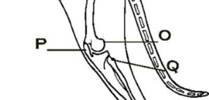
- Distal femoral epiphyseal (O) closes between 4.5 and 13 months.
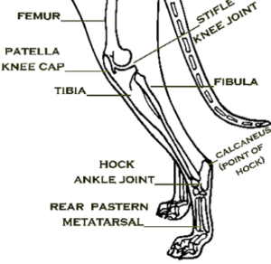
STIFLE / KNEE - Hind/Back
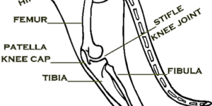
The dog’s stifle joint consists of three primary bones:
- Femur: The upper leg bone. Distal femoral epiphyseal (O) closes between 4.5 and 13 months.
- Tibia: The lower leg bone. The tibial condyle (P) closes between 4.7 and 13 months.
- Patella: Also known as the kneecap, it protects the front of the joint and aids in leg extension.
- Additionally, there are small sesamoid bones called fabellae located in the stifle region, which assist in muscle function.
HOCK and Hind PAW
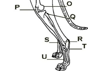
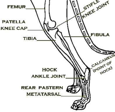
The dog’s hock joint consists of several key bones.
- Tibia: The main shin bone that connects to the hock. Distal tibial epiphyseal (S) closes between 4.5 and 16.5 months.
- Fibula: A smaller bone located alongside the tibia. Distal fibular epiphyseal (R) closes between 4.5 and 16.5 months.
- Tarsal bones: There are seven tarsal bones that make up the hock. Tarsal bones (T) closes around 3-8 months.
These bones include the:
- Talus: The bone that articulates with the tibia.
- Calcaneus: The heel bone, which is the largest tarsal bone.
- Navicular: Located in front of the talus.
- Cuneiforms: Three bones (medial, intermediate, and lateral) that are positioned in front of the navicular.
- Metatarsal bones: Five long bones that extend from the tarsals to the toes, although they are not part of the hock itself, they connect to it. Metatarsal bones (U) close between 6 and 9 months.
Hind Paw:
- Metatarsal closes between 6.7 and 9 months.
- Phalanx closes between 5.3 and 7 months.

CARPUS / WRIST - Front
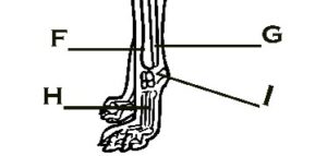
The wrist and paw bones close early around 3-9 months, but some continue growing up to 17 months, affecting stability.
- Radial (carpal/bottom – F) closes between 4.5 and 17 months
- Ulna (carpal/bottom – G) closes between 7.2-15
- Wrist – Carpals (I) closes around 3.3 months
- Metacarpals (H) closes between 6.7 and 9 months.
- The wrist closes at around 3.3 months while the bones above it continue growing for up to another 14 months, to provide stability at the wrist during weight bearing.
WRIST / PAW - Front
The little bones in the paw take 5 to 9 months to close, and there is the dewclaw (puppy’s thumb) that closes between 5.3 and 7 months of age.
- Wrist – Carpals (I) closes around 3.3 months.
- Metacarpals (H) closes between 6.7 and 9 months.
- Dewclaw (thumb) closes between 5.3 and 7 months.
- Phalanx closes between 5.3 and 7 months.
HOCK and Hind PAW
The dog’s hock joint consists of several key bones.
- Tibia: The main shin bone that connects to the hock. Distal tibial epiphyseal (S) closes between 4.5 and 16.5 months.
- Fibula: A smaller bone located alongside the tibia. Distal fibular epiphyseal (R) closes between 4.5 and 16.5 months.
- Tarsal bones: There are seven tarsal bones that make up the hock. Tarsal bones (T) closes around 3-8 months.
These bones include the:
- Talus: The bone that articulates with the tibia.
- Calcaneus: The heel bone, which is the largest tarsal bone.
- Navicular: Located in front of the talus.
- Cuneiforms: Three bones (medial, intermediate, and lateral) that are positioned in front of the navicular.
- Metatarsal bones: Five long bones that extend from the tarsals to the toes, although they are not part of the hock itself, they connect to it. Metatarsal bones (U) close between 6 and 9 months.
Hind Paw:
- Metatarsal closes between 6.7 and 9 months.
- Phalanx closes between 5.3 and 7 months.